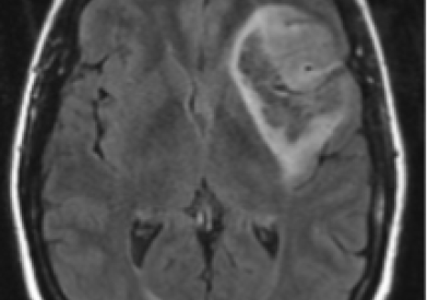
Craniopharyngioma

  • Dana-Farber/Brigham and Women's Cancer Care

    Craniopharyngioma is a benign brain tumor that may be considered malignant because it can damage the hypothalamus, the area of the brain that controls body temperature, hunger and thirst. Learn about craniopharyngioma and find information on how we support and care for people with craniopharyngioma before, during, and after treatment.

Treatment 

The Center for Neuro-Oncology offers state-of the-art treatments for patients with brain tumors, spinal cord tumors, and neurologic complications from cancer.

We specialize in services for patients with benign or malignant tumors, including:

  • Brain tumors: high-grade glioma, astrocytoma, oligodendroglioma, anaplastic glioma, glioblastoma, medulloblastoma, and meningioma
  • Pituitary, acoustic and skull base tumors: chordoma, meningioma, acoustic neuroma, hemangioblastoma, and pituitary adenoma

Our clinicians are experts in precise neurosurgical techniques, radiation therapy that targets tumors while sparing surrounding tissues, and a variety of chemotherapy and molecular drugs targeting your type of cancer.

Our approach to compassionate care centers on the needs of our patients and their families. Our specialists work as a team to ensure that you receive carefully coordinated treatment and support. We focus on helping you cope with your illness and maintain the quality of your life.

Our research program is a leader in clinical trials for brain tumors. Our goal is to bring laboratory discoveries directly to patients, allowing us to offer new treatment options. We offer our patients access to a range of clinical trials for various types and stages of brain and spinal cord cancers.

We believe that you and your family are key members of the care team. While we understand your illness, you understand your own body. We welcome your questions and value your participation as we make decisions together.

Learn more about treatment for brain tumors in the Center for Neuro-oncology 

Contact us 

New patients 

If you have never been seen before at Dana-Farber/Brigham and Women's Cancer Center, please call 877-442-3324 or use this online form to request an appointment.

For all other inquiries, please call 617-632-2166 

Referring physicians: 617-632-3703 

Fax: 617-632-4773 

Information for: Patients | Healthcare Professionals

General Information About Adult Brain Tumors

An adult brain tumor is a disease in which abnormal cells form in the tissues of the brain.

There are many types of brain and spinal cordtumors. The tumors are formed by the abnormal growth of cells and may begin in different parts of the brain or spinal cord. Together, the brain and spinal cord make up the central nervous system (CNS).

The tumors may be benign (not cancer) or malignant (cancer). Benign brain tumors grow and press on nearby areas of the brain. They rarely spread into other tissues and may recur (come back). Malignant brain tumors are likely to grow quickly and spread into other brain tissue. When a tumor grows into or presses on an area of the brain, it may keep that part of the brain from working the way it should. Both benign and malignant brain tumors can cause symptoms and, sometimes, death.

Brain tumors can occur in both adults and children. However, treatment for children may be different than treatment for adults. (See the PDQ summary on Childhood Brain and Spinal Cord Tumors Treatment Overview for more information on the treatment of children.)

For information about lymphoma that begins in the brain, see the PDQ summary on Primary CNS Lymphoma Treatment.

A brain tumor that starts in another part of the body and spreads to the brain is called a metastatic tumor.

Tumors that start in the brain are called primary brain tumors. Often, tumors found in the brain have started somewhere else in the body and spread to one or more parts of the brain. These are called metastatic brain tumors (or brain metastases). Metastatic brain tumors are more common than primary brain tumors.

The types of cancer that commonly spread to the brain are melanoma and cancer of the breast, colon, lung, and unknown primary site. The types of cancer that commonly spread to the spinal cord are lymphoma and cancer of the lung, breast, and prostate. About half of metastatic brain and spinal cord tumors are caused by lung cancer. Leukemia, lymphoma, breast cancer, and gastrointestinal cancer may spread to the leptomeninges (the two innermost membranes covering the brain and spinal cord).

See the following PDQ summaries for more information on cancers that commonly spread to the brain and spinal cord:

  • Adult Hodgkin Lymphoma Treatment
  • Adult Non-Hodgkin Lymphoma Treatment
  • Breast Cancer Treatment
  • Carcinoma of Unknown Primary Treatment
  • Colon Cancer Treatment
  • Melanoma Treatment
  • Non-Small Cell Lung Cancer Treatment
  • Small Cell Lung Cancer Treatment

The brain controls many important body functions.

The brain has three major parts:

  • The cerebrum is the largest part of the brain. It is at the top of the head. The cerebrum controls thinking, learning, problem solving, emotions, speech, reading, writing, and voluntary movement.
  • The cerebellum is in the lower back of the brain (near the middle of the back of the head). It controls movement, balance, and posture.
  • The brain stem connects the brain to the spinal cord. It is in the lowest part of the brain (just above the back of the neck). The brain stem controls breathing, heart rate, and the nerves and muscles used in seeing, hearing, walking, talking, and eating.

The spinal cord connects the brain to nerves in most parts of the body.

The spinal cord is a column of nerve tissue that runs from the brain stem down the center of the back. It is covered by three thin layers of tissue called membranes. These membranes are surrounded by the vertebrae (back bones). Spinal cord nerves carry messages between the brain and the rest of the body, such as a signal from the brain to cause muscles to move or from the skin to the brain about the sense of touch.

There are different types of brain and spinal cord tumors.

Brain and spinal cord tumors are named based on the type of cell they formed in and where the tumor first formed in the CNS. The grade of a tumor may be used to tell the difference between slow- and fast-growing types of the tumor. The grade of a tumor is based on how abnormal the cancer cells look under a microscope and how quickly the tumor is likely to grow and spread.

Tumor Grading System

  • Grade I (low-grade) — The tumor grows slowly, has cells that look a lot like normal cells, and rarely spreads into nearby tissues. It may be possible to remove the entire tumor by surgery.
  • Grade II — The tumor grows slowly, but may spread into nearby tissue and may recur (come back). Some tumors may become a higher-grade tumor.
  • Grade III — The tumor grows quickly, is likely to spread into nearby tissue, and the tumor cells look very different from normal cells.
  • Grade IV (high-grade) — The tumor grows and spreads very quickly and the cells do not look like normal cells. There may be areas of dead cells in the tumor. Grade IV brain tumors are harder to cure than lower-grade tumors.

Astrocytic Tumors

An astrocytic tumor begins in star-shaped brain cells called astrocytes, which help keep nerve cells healthy. An astrocyte is a type of glial cell and is sometimes called a glioma. Astrocytic tumors include the following:

  • Brain stem glioma: A brain stem glioma forms in the brain stem, which is the part of the brain connected to the spinal cord. It is often a high-grade tumor, which spreads widely through the brain stem and is hard to cure. A brain stem glioma rarely occurs in adults. (See the PDQ summary on Childhood Brain Stem Glioma Treatment for more information.)
  • Pineal astrocytic tumor: A pineal astrocytic tumor forms in tissue around the pineal gland and may be any grade. The pineal gland is a tiny organ in the brain that makes melatonin, a hormone that helps control the sleeping and waking cycle.
  • Pilocytic astrocytoma (grade I): A pilocyticastrocytoma grows slowly in the brain or spinal cord. It may be in the form of a cyst and rarely spreads into nearby tissues. This type of tumor is most common in children and young adults and in people with neurofibromatosis type 1 (NF1). A pilocytic astrocytoma rarely causes death.
  • Diffuse astrocytoma (grade II): A diffuse astrocytoma grows slowly, but often spreads into nearby tissues. Sometimes a diffuse astrocytoma progresses to a higher grade and becomes an anaplastic astrocytoma or a glioblastoma. A diffuse astrocytoma can form in any part of the brain but most often forms in the cerebrum. It is most common in young adults and in people with Li-Fraumeni syndrome. It is also called a low-grade diffuse astrocytoma.
  • Anaplastic astrocytoma (grade III): An anaplastic astrocytoma grows quickly and spreads into nearby tissues. An anaplastic astrocytoma may progress to a higher grade and become a glioblastoma. An anaplastic astrocytoma forms most often in the cerebrum and is most common in adults. An anaplastic astrocytoma is also called a malignant astrocytoma or high-grade astrocytoma.
  • Glioblastoma (grade IV): A glioblastoma grows and spreads very quickly. A glioblastoma forms most often in the cerebrum. This type of tumor is most common in adults. This type of tumor has a poor prognosis. It is also called glioblastoma multiforme.

Oligodendroglial Tumors

An oligodendroglial tumor begins in brain cells called oligodendrocytes, which help keep nerve cells healthy. Oligodendrocytes are a type of glial cell and are sometimes called a glioma. Grades of oligodendroglial tumors include the following:

  • Oligodendroglioma (grade II): An oligodendroglioma grows and spreads slowly and the tumor cells look very much like normal cells. This type of tumor most often forms in the cerebrum. An oligodendroglioma is most common in adults.
  • Anaplastic oligodendroglioma (grade III): An anaplastic oligodendroglioma grows quickly and the tumor cells look very different from normal cells. It may grow in one place or in many places throughout the brain. This type of cancer most often forms in the cerebrum.

Mixed Gliomas

A mixed glioma is a brain tumor that has two types of tumor cells in it — oligodendrocytes and astrocytes. This type of tumor most often forms in the cerebrum.

  • Oligoastrocytoma (grade II): An oligoastrocytoma is a slow-growing tumor and the tumor cells don't look very different from normal cells.
  • Anaplastic oligoastrocytoma (grade III): The tumor cells in an anaplastic oligoastrocytoma look very different from normal cells.

Ependymal Tumors

An ependymal tumor usually begins in cells that line the fluid-filled spaces in the brain and around the spinal cord. Ependymal cells are a type of glial cell and are sometimes called a glioma. Grades of ependymal tumors include the following:

  • Ependymoma (grade I or II): A grade I or II ependymoma grows slowly and has cells that look very much like normal cells. There are two types of grade I ependymoma — myxopapillary ependymoma and subependymoma. These tumors are most common in adults. A grade II ependymoma grows in the ventricle and its connecting paths or in the spinal cord. It is most common in children and young adults and in people with neurofibromatosis type 2 (NF2). (See the PDQ summary on Childhood Ependymoma Treatment for more information.)
  • Anaplastic ependymoma (grade III): An anaplastic ependymoma grows very quickly and has a poor prognosis.

Embryonal Cell Tumors: Medulloblastoma (Grade IV)

A medulloblastoma is a type of embryonal tumor. The tumor forms in brain cells when the fetus is beginning to develop. This type of brain tumor often begins in the cerebellum. The tumor may spread from the brain to the spine through the cerebrospinal fluid (CSF). A medulloblastoma occurs most often in children or young adults and in people with Turcot syndrome type 2 or nevoid basal cell carcinoma syndrome.

See the following PDQ summaries for more information on embryonal tumors in children:

  • Childhood Central Nervous System Embryonal Tumors Treatment
  • Childhood Central Nervous System Atypical Teratoid/Rhabdoid Tumor Treatment

Pineal Parenchymal Tumors

A pineal parenchymal tumor forms in parenchymal cells or pineocytes, which are the cells that make up most of the pineal gland. These tumors are different from pineal astrocytic tumors. Grades of pineal parenchymal tumors include the following:

  • Pineocytomas (grade II): A pineocytoma is a slow-growing pineal tumor that occurs most often in adults.
  • Pineoblastomas (grade IV): A pineoblastoma is a rare tumor that is very likely to spread. This type of tumor is most common in children. (See the PDQ summary on Childhood Central Nervous System Embryonal Tumors Treatment for more information.)

Meningeal Tumors

A meningeal tumor, also called a meningioma, forms in the meninges (thin layers of tissue that cover the brain and spinal cord). It can form from different types of brain or spinal cord cells. A meningioma is most common in adults. Types of meningeal tumors include the following:

  • Meningioma (grade I): A grade I meningioma is the most common type of meningeal tumor. A grade I meningioma is a slow-growing, benign tumor that forms most often in the dura mater (the layer of tissue that covers the brain and is closest to the skull). It is most common in women.
  • Meningioma (grade II and III): This is a rare, malignant meningeal tumor. It grows quickly and is likely to spread within the brain and spinal cord. A grade III meningioma is most common in men.

A hemangiopericytoma is not a meningeal tumor but is treated like a grade II or III meningioma. A hemangiopericytoma usually forms in the dura mater. It often recurs (comes back) after treatment and usually spreads to other parts of the body.

Germ Cell Tumors

A germ cell tumor forms in germ cells, which are the cells that develop into sperm in men or ova (eggs) in women. Germ cell tumors usually form in the center of the brain, near the pineal gland. Germ cell tumors can spread to other parts of the brain and spinal cord. There are different types of germ cell tumors. These include germinomas, teratomas, embryonal yolk sac carcinomas, and choriocarcinomas. Germ cell tumors can be either benign or malignant.

Most germ cell tumors occur in children and in people with Klinefelter syndrome. (See the PDQ summary on Childhood Brain and Spinal Cord Tumors Treatment Overview for more information.)

Tumors of the Sellar Region: Craniopharyngioma (Grade I) and Pituitary Tumor

A tumor of the sellar region begins in the center of the brain, just above the back of the nose. It can form from different types of brain or spinal cord cells.

  • Craniopharyngioma (grade I): A craniopharyngioma is a rare tumor that usually forms just above the pituitary gland (a pea-sized organ at the bottom of the brain that controls other glands). The tumor may grow into nearby tissues, including the pituitary gland and optic nerves. This can affect many functions, including hormone-making and vision. These tumors occur in adults and children. (See the PDQ summary on Childhood Craniopharyngioma Treatment for more information on the treatment of children.)
  • Pituitary tumor: See the PDQ summary on Pituitary Tumors Treatment for more information.

Other Adult Brain Tumors

There are many other types of adult brain tumors that are rare and are not discussed in this summary. See the PDQ health professional summary on Adult Brain Tumors Treatment for information about these other types of adult brain tumors:

  • Pleomorphic xanthoastrocytoma
  • Subependymal giant cell astrocytoma
  • Astroblastoma
  • Chordoid glioma of the third ventricle
  • Gliomatosis cerebri
  • Gangliocytoma or ganglioglioma
  • Desmoplastic infantile astrocytoma or ganglioglioma
  • Dysembryoplastic neuroepithelial tumor
  • Central neurocytoma
  • Cerebellar liponeurocytoma
  • Paraganglioma
  • Ependymoblastoma
  • Supratentorial primitive neuroectodermal tumor (PNET)
  • Choroid plexus papilloma or carcinoma
  • Pineal parenchymal tumors of intermediate differentiation
  • Melanocytic lesions
  • Capillary hemangioblastoma
  • Schwannoma

Recurrent Brain Tumors

A recurrent brain tumor is a tumor that has recurred (come back) after it has been treated. Brain tumors often recur, sometimes many years after the first tumor. The tumor may recur at the same place in the brain or in other parts of the central nervous system.

The cause of most adult brain tumors is unknown.

Anything that increases your chance of getting a disease is called a risk factor. Having a risk factor does not mean that you will get cancer; not having risk factors doesn’t mean that you will not get cancer. People who think they may be at risk should discuss this with their doctor. There are few known risk factors for brain tumors. The following conditions may increase the risk of developing certain types of brain tumors:

  • Being exposed to vinyl chloride may increase the risk of glioma.
  • Past treatment with radiation therapy to the scalp or brain may increase the risk of meningioma.
  • Infection with the Epstein-Barr virus, having AIDS (acquired immunodeficiency syndrome), or receiving an organ transplant may increase the risk of primary CNS lymphoma. (See the PDQ summary on Primary CNS Lymphoma for more information.)
  • Having certain genetic syndromes may increase the risk of developing the following types of brain tumors:
    • Neurofibromatosis type 1 or 2.
    • von Hippel-Lindau disease.
    • Tuberous sclerosis.
    • Li-Fraumeni syndrome.
    • Turcot syndrome type 1 and type 2.
    • Klinefelter syndrome.
    • Nevoid basal cell carcinoma syndrome.
     

The symptoms of adult brain and spinal cord tumors are not the same in every person.

The symptoms caused by a brain tumor depend on where the tumor formed in the brain, the functions controlled by that part of the brain, and the size of the tumor. Headaches and other symptoms may be caused by adult brain tumors. Other conditions may cause the same symptoms. A doctor should be consulted if any of the following problems occur:

Brain Tumors

  • Morning headache or headache that goes away after vomiting.
  • Frequent nausea and vomiting.
  • Vision, hearing, and speech problems.
  • Loss of balance and trouble walking.
  • Weakness on one side of the body.
  • Unusual sleepiness or change in activity level.
  • Unusual changes in personality or behavior.
  • Seizures.

Spinal Cord Tumors

  • Back pain or pain that spreads from the back towards the arms or legs.
  • A change in bowel habits or trouble urinating.
  • Weakness in the legs.
  • Trouble walking.

Tests that examine the brain and spinal cord are used to detect (find) adult brain tumors.

The following tests and procedures may be used:

  • Physical exam and history: An exam of the body to check general signs of health, including checking for signs of disease, such as lumps or anything else that seems unusual. A history of the patient’s health habits and past illnesses and treatments will also be taken.
  • Neurological exam: A series of questions and tests to check the brain, spinal cord, and nerve function. The exam checks a person’s mental status, coordination, and ability to walk normally, and how well the muscles, senses, and reflexes work. This may also be called a neuro exam or a neurologic exam.
  • Visual field exam: An exam to check a person’s field of vision (the total area in which objects can be seen). This test measures both central vision (how much a person can see when looking straight ahead) and peripheral vision (how much a person can see in all other directions while staring straight ahead). Any loss of vision may be a sign of a tumor that has damaged or pressed on the parts of the brain that affect eyesight.
  • Tumor marker test: A procedure in which a sample of blood, urine, or tissue is checked to measure the amounts of certain substances made by organs, tissues, or tumor cells in the body. Certain substances are linked to specific types of cancer when found in increased levels in the body. These are called tumor markers.
  • Gene testing: A laboratory test in which a sample of blood or tissue is tested for changes in a chromosome that has been linked with a certain type of brain tumor.
  • Lumbar puncture: A procedure used to collect cerebrospinal fluid from the spinal column. This is done by placing a needle into the spinal column. The cerebrospinal fluid is viewed under a microscope by a pathologist to check for signs of cancer. This procedure is also called an LP or spinal tap.
  • CT scan (CAT scan): A procedure that makes a series of detailed pictures of areas inside the body, taken from different angles. The pictures are made by a computer linked to an x-ray machine. A dye may be injected into a vein or swallowed to help the organs or tissues show up more clearly. This procedure is also called computed tomography, computerized tomography, or computerized axial tomography.
  • MRI (magnetic resonance imaging) with gadolinium: A procedure that uses a magnet, radio waves, and a computer to make a series of detailed pictures of the brain and spinal cord. A substance called gadolinium is injected into a vein. The gadolinium collects around the cancer cells so they show up brighter in the picture. This procedure is also called nuclear magnetic resonance imaging (NMRI). Sometimes a procedure called magnetic resonance spectroscopy (MRS) is done during the MRI scan. An MRS is used to diagnose tumors, based on their chemical make-up.
  • SPECT scan (single photon emission computed tomography scan): A procedure that uses a special camera linked to a computer to make a 3-dimensional (3-D) picture of the brain. A small amount of a radioactive substance is injected into a vein or inhaled through the nose. As the substance travels through the blood, the camera rotates around the head and takes pictures of the brain. There will be increased blood flow and more chemical reactions (metabolism) in areas where cancer cells are growing. These areas will show up brighter in the picture. This procedure may be done just before or after a CT scan.
  • PET scan (positron emission tomography scan): A procedure to find malignant tumor cells in the body. A small amount of radioactive glucose (sugar) is injected into a vein. The PET scanner rotates around the body and makes a picture of where glucose is being used in the brain. Malignant tumor cells show up brighter in the picture because they are more active and take up more glucose than normal cells do.
  • Angiogram: A procedure to look at blood vessels and the flow of blood in the brain. A contrast dye is injected into the blood vessel. As the contrast dye moves through the blood vessel, x-rays are taken to see if there are any blockages.

Most adult brain tumors are diagnosed and removed in surgery.

If doctors think there may be a brain tumor, a biopsy may be done to remove a sample of tissue. For tumors in the brain, the biopsy is done by removing part of the skull and using a needle to remove the tissue sample. A pathologist views the tissue under a microscope to look for cancer cells. If cancer cells are found, the doctor may remove as much tumor as safely possible during the same surgery. After the surgery, a pathologist checks the cancer cells to find out the type and grade of brain tumor. The grade of the tumor is based on how abnormal the cancer cells look under a microscope and how quickly the tumor is likely to grow and spread. A CT scan or MRI may be used to find out if any cancer cells remain after surgery.

The following tests may be done on the tumor tissue that is removed:

  • Immunohistochemistry study: A laboratory test in which a substance such as an antibody, dye, or radioisotope is added to a sample of cancer tissue to test for certain antigens. This type of study is used to tell the difference between different types of cancer.
  • Light and electron microscopy: A laboratory test in which cells in a sample of tissue are viewed under regular and high-powered microscopes to look for certain changes in the cells.
  • Cytogenetic analysis: A laboratory test in which cells in a sample of tissue are viewed under a microscope to look for certain changes in the chromosomes.

Sometimes a biopsy or surgery cannot be done safely because of where the tumor formed in the brain or spinal cord. These tumors are diagnosed based on the results of imaging tests and other procedures.

Certain factors affect prognosis (chance of recovery) and treatment options.

The prognosis (chance of recovery) and treatment options for primary brain tumors depend on the following:

  • The type and grade of the tumor.
  • Where the tumor is in the brain.
  • Whether the tumor can be removed by surgery.
  • Whether cancer cells remain after surgery.
  • Whether there are certain changes in the chromosomes.
  • Whether the cancer has just been diagnosed or has recurred (come back).
  • The patient's general health.

The prognosis and treatment options for metastatic brain tumors depend on the following:

  • Whether the patient is younger than 60 years.
  • Whether there are more than two tumors in the brain or spinal cord.
  • Where in the brain or spinal cord the tumors are.
  • How well the tumor responds to treatment.
  • Whether the primary tumor continues to grow or spread.

The prognosis is better for brain metastases from breast cancer than from other types of primary cancer. The prognosis is worse for brain metastases from colon cancer.

Stages of Adult Brain Tumors

There is no standard staging system for adult brain tumors.

The extent or spread of cancer is usually described as stages. There is no standard staging system for brain tumors. Brain tumors that begin in the brain may spread to other parts of the brain and spinal cord, but they rarely spread to other parts of the body. Treatment of brain tumors is based on the type of cell in which the tumor began, where the tumor formed in the central nervous system, the amount of cancer left after surgery, and the grade of the tumor. Treatment of brain tumors that have spread to the brain from other parts of the body is based on the number of tumors in the brain.

Imaging tests may be repeated after surgery to help plan more treatment.

After the tumor is removed in surgery, some of the tests and procedures used to detect the brain tumor may be repeated to find out how much tumor is left.

Treatment Option Overview

There are different types of treatment for patients with adult brain tumors.

Different types of treatment are available for patients with adult brain tumors. Some treatments are standard (the currently used treatment), and some are being tested in clinical trials. A treatment clinical trial is a research study meant to help improve current treatments or obtain information on new treatments for patients with cancer. When clinical trials show that a new treatment is better than the standard treatment, the new treatment may become the standard treatment. Patients may want to think about taking part in a clinical trial. Some clinical trials are open only to patients who have not started treatment.

Three types of standard treatment are used:

Surgery

Surgery is used to diagnose and treat adult brain tumors, as described in the General Information section of this summary.

Even if the doctor removes all the cancer that can be seen at the time of the surgery, some patients may be given chemotherapy or radiation therapy after surgery to kill any cancer cells that are left. Treatment given after the surgery, to lower the risk that the cancer will come back, is called adjuvant therapy.

Radiation therapy

Radiation therapy is a cancer treatment that uses high-energy x-rays or other types of radiation to kill cancer cells or keep them from growing. There are two types of radiation therapy. External radiation therapy uses a machine outside the body to send radiation toward the cancer. One type of external radiation therapy is hyperfractionated radiation therapy, in which the total dose of radiation is divided into small doses given more than once a day. Internal radiation therapy uses a radioactive substance sealed in needles, seeds, wires, or catheters that are placed directly into or near the cancer. The way the radiation therapy is given depends on the type of tumor and where it is in the brain.

Chemotherapy

Chemotherapy is a cancer treatment that uses drugs to stop the growth of cancer cells, either by killing the cells or by stopping them from dividing. When chemotherapy is taken by mouth or injected into a vein or muscle, the drugs enter the bloodstream and can reach cancer cells throughout the body (systemic chemotherapy). When chemotherapy is placed directly into the spinal column, an organ, or a body cavity such as the abdomen, the drugs mainly affect cancer cells in those areas (regional chemotherapy). Combination chemotherapy is treatment using more than one anticancer drug. To treat brain tumors, a dissolving wafer may be used to deliver an anticancer drug directly to the brain tumor site after the tumor has been removed by surgery. The way the chemotherapy is given depends on the type of tumor and where it is in the brain.

New types of treatment are being tested in clinical trials.

This summary section refers to specific treatments under study in clinical trials, but it may not mention every new treatment being studied. Information about clinical trials is available from the NCI Web site.

Other types of radiation therapy

  • Radiosensitizers: Drugs that make tumor cells more sensitive to radiation therapy. Combining radiation therapy with radiosensitizers may kill more tumor cells.
  • Hyperfractionated radiation therapy: Hyperfractionated radiation therapy is radiation treatment in which the total dose of radiation is divided into small doses and the treatments are given more than once a day. Hyperfractionated radiation therapy is used as a standard treatment for a brain stem glioma.
  • Accelerated-fraction radiation therapy: Radiation treatment in which the total dose of radiation is divided into small doses and the treatments are given more than once a day. The total dose of radiation is also given over a shorter period of time (fewer days) compared to standard radiation therapy.
  • Intraoperative radiation therapy: A cancer treatment that uses high-energy x-rays to kill cancer cells during cancer surgery.
  • Stereotactic radiosurgery: A type of radiation therapy that uses a rigid head frame attached to the skull to aim high-dose radiation beams directly at the tumors. This causes less damage to nearby healthy tissue. This is also called stereotaxic radiosurgery and radiation surgery. This procedure does not involve surgery.

Hyperthermia therapy

Hyperthermia therapy is a treatment in which body tissue is heated above normal temperature to damage and kill cancer cells or to make cancer cells more sensitive to the effects of radiation and certain anticancer drugs.

Biologic therapy

Biologic therapy is a treatment that uses the patient’s immune system to fight cancer. Substances made by the body or made in a laboratory are used to boost, direct, or restore the body’s natural defenses against cancer. This type of cancer treatment is also called biotherapy or immunotherapy.

Patients may want to think about taking part in a clinical trial.

For some patients, taking part in a clinical trial may be the best treatment choice. Clinical trials are part of the cancer research process. Clinical trials are done to find out if new cancer treatments are safe and effective or better than the standard treatment.

Many of today's standard treatments for cancer are based on earlier clinical trials. Patients who take part in a clinical trial may receive the standard treatment or be among the first to receive a new treatment.

Patients who take part in clinical trials also help improve the way cancer will be treated in the future. Even when clinical trials do not lead to effective new treatments, they often answer important questions and help move research forward.

Patients can enter clinical trials before, during, or after starting their cancer treatment.

Some clinical trials only include patients who have not yet received treatment. Other trials test treatments for patients whose cancer has not gotten better. There are also clinical trials that test new ways to stop cancer from recurring (coming back) or reduce the side effects of cancer treatment.

Clinical trials are taking place in many parts of the country. See the Treatment Options section that follows for links to current treatment clinical trials. These have been retrieved from NCI's clinical trials database.

Follow-up tests may be needed.

Some of the tests that were done to diagnose the cancer or to find out the stage of the cancer may be repeated. Some tests will be repeated in order to see how well the treatment is working. Decisions about whether to continue, change, or stop treatment may be based on the results of these tests. This is sometimes called re-staging.

Some of the tests will continue to be done from time to time after treatment has ended. The results of these tests can show if your condition has changed or if the cancer has recurred (come back). These tests are sometimes called follow-up tests or check-ups.

Treatment Options by Type of Adult Brain Tumor

A link to a list of current clinical trials is included for each treatment section. For some types or stages of cancer, there may not be any trials listed. Check with your doctor for clinical trials that are not listed here but may be right for you.

Astrocytic Tumors

Brain Stem Gliomas

Treatment of brain stem gliomas is radiation therapy, including hyperfractionated radiation therapy.

Check for U.S. clinical trials from NCI's PDQ Cancer Clinical Trials Registry that are now accepting patients with adult brain stem glioma. For more specific results, refine the search by using other search features, such as the location of the trial, the type of treatment, or the name of the drug. General information about clinical trials is available from the NCI Web site.

Pineal Astrocytic Tumors

Treatment of pinealastrocytictumors may include the following:

  • Surgery and radiation therapy, with or without chemotherapy.
  • A clinical trial of external radiation therapy with or without radiosensitizers, intraoperative radiation therapy, or hyperthermia therapy.
  • A clinical trial of external radiation therapy, followed by new anticancer drugs and biologic therapy.

Check for U.S. clinical trials from NCI's PDQ Cancer Clinical Trials Registry that are now accepting patients with adult pineal gland astrocytoma. For more specific results, refine the search by using other search features, such as the location of the trial, the type of treatment, or the name of the drug. General information about clinical trials is available from the NCI Web site.

Pilocytic Astrocytomas

Treatment of pilocyticastrocytoma is usually surgery with or without radiation therapy.

Check for U.S. clinical trials from NCI's PDQ Cancer Clinical Trials Registry that are now accepting patients with adult pilocytic astrocytoma. For more specific results, refine the search by using other search features, such as the location of the trial, the type of treatment, or the name of the drug. General information about clinical trials is available from the NCI Web site.

Diffuse Astrocytomas

Treatment of diffuseastrocytoma may include the following:

  • Surgery with or without radiation therapy.
  • A clinical trial of radiation therapy with or without chemotherapy for tumors that could not be completely removed by surgery.
  • A clinical trial of radiation therapy given when the tumor progresses.
  • A clinical trial to compare high-dose and low-dose radiation therapy.

Check for U.S. clinical trials from NCI's PDQ Cancer Clinical Trials Registry that are now accepting patients with adult diffuse astrocytoma. For more specific results, refine the search by using other search features, such as the location of the trial, the type of treatment, or the name of the drug. General information about clinical trials is available from the NCI Web site.

Anaplastic Astrocytomas

Treatment of anaplasticastrocytoma may include the following:

  • Surgery and radiation therapy, with or without chemotherapy.
  • A clinical trial of external radiation therapy and one of the following:
    • Hyperfractionated radiation therapy.
    • Accelerated-fraction radiation therapy.
    • Stereotactic radiosurgery.
    • Radiosensitizers.
    • Hyperthermia therapy.
    • Internal radiation therapy.
    • Intraoperative radiation therapy.
     
  • A clinical trial of external radiation therapy, followed by new anticancer drugs and biologic therapy.
  • A clinical trial of chemotherapy combined with hyperfractionated radiation therapy or internal and external radiation therapy.
  • A clinical trial of chemotherapy placed into the brain during surgery.

Check for U.S. clinical trials from NCI's PDQ Cancer Clinical Trials Registry that are now accepting patients with adult anaplastic astrocytoma. For more specific results, refine the search by using other search features, such as the location of the trial, the type of treatment, or the name of the drug. General information about clinical trials is available from the NCI Web site.

Glioblastoma

Treatment of glioblastoma may include the following:

  • Surgery and radiation therapy, with or without chemotherapy.
  • Chemotherapy placed into the brain during surgery.
  • Radiation therapy given at the same time as chemotherapy.
  • A clinical trial of external radiation therapy and one of the following:
    • Hyperfractionated radiation therapy.
    • Accelerated-fraction radiation therapy.
    • Stereotactic radiosurgery.
    • Radiosensitizers.
    • Hyperthermia therapy.
    • Internal radiation therapy.
    • Intraoperative radiation therapy.
     
  • A clinical trial of external radiation therapy, followed by new anticancer drugs and biologic therapy.
  • A clinical trial of a new treatment.

Check for U.S. clinical trials from NCI's PDQ Cancer Clinical Trials Registry that are now accepting patients with adult glioblastoma. For more specific results, refine the search by using other search features, such as the location of the trial, the type of treatment, or the name of the drug. General information about clinical trials is available from the NCI Web site.

Oligodendroglial Tumors

Treatment of oligodendrogliomas may include the following:

  • Surgery with or without radiation therapy.
  • Chemotherapy.
  • A clinical trial of radiation therapy with or without chemotherapy, for tumors that could not be completely removed by surgery.
  • A clinical trial of chemotherapy.

Treatment of anaplastic oligodendroglioma may include the following:

  • Surgery and radiation therapy, with or without chemotherapy.
  • Combination chemotherapy.
  • Radiation therapy with or without combination chemotherapy.
  • A clinical trial of a new treatment.

Check for U.S. clinical trials from NCI's PDQ Cancer Clinical Trials Registry that are now accepting patients with adult oligodendroglial tumors. For more specific results, refine the search by using other search features, such as the location of the trial, the type of treatment, or the name of the drug. General information about clinical trials is available from the NCI Web site.

Mixed Gliomas

Treatment of mixed gliomas may include the following:

  • Surgery and radiation therapy, with or without chemotherapy.
  • Chemotherapy.
  • A clinical trial of external radiation therapy and one of the following:
    • Internal radiation therapy.
    • Radiosensitizers.
    • Hyperthermia therapy.
    • Intraoperative radiation therapy.
     
  • A clinical trial of external radiation therapy, followed by new anticancer drugs and biologic therapy.

Check for U.S. clinical trials from NCI's PDQ Cancer Clinical Trials Registry that are now accepting patients with adult mixed glioma. For more specific results, refine the search by using other search features, such as the location of the trial, the type of treatment, or the name of the drug. General information about clinical trials is available from the NCI Web site.

Ependymal Tumors

Treatment of grade I and grade II ependymomas is usually surgery with or without radiation therapy.

Treatment of grade III anaplastic ependymoma may include the following:

  • Surgery and radiation therapy.
  • A clinical trial of chemotherapy before, during, or after radiation therapy.

Check for U.S. clinical trials from NCI's PDQ Cancer Clinical Trials Registry that are now accepting patients with adult ependymal tumors. For more specific results, refine the search by using other search features, such as the location of the trial, the type of treatment, or the name of the drug. General information about clinical trials is available from the NCI Web site.

Embryonal Cell Tumors: Medulloblastoma

Treatment of medulloblastomas may include the following:

  • Surgery and radiation therapy to the brain and spine.
  • Chemotherapy.
  • A clinical trial of surgery and radiation therapy to the brain and spine.
  • A clinical trial of chemotherapy.

Check for U.S. clinical trials from NCI's PDQ Cancer Clinical Trials Registry that are now accepting patients with adult medulloblastoma. For more specific results, refine the search by using other search features, such as the location of the trial, the type of treatment, or the name of the drug. General information about clinical trials is available from the NCI Web site.

Pineal Parenchymal Tumors

Treatment of pinealparenchymaltumors may include the following:

  • Surgery and radiation therapy, for pineocytomas.
  • Surgery, radiation therapy, and chemotherapy, for pineoblastomas.
  • A clinical trial of external radiation therapy and one of the following:
    • Radiosensitizers.
    • Hyperthermia therapy.
    • Intraoperative radiation therapy.
     
  • A clinical trial of external radiation therapy, followed by new anticancer drugs and biologic therapy.

Check for U.S. clinical trials from NCI's PDQ Cancer Clinical Trials Registry that are now accepting patients with adult pineal parenchymal tumor. For more specific results, refine the search by using other search features, such as the location of the trial, the type of treatment, or the name of the drug. General information about clinical trials is available from the NCI Web site.

Meningeal Tumors

Treatment of grade I meningiomas may include the following:

  • Surgery with or without radiation therapy.
  • Radiation therapy for tumors that cannot be removed by surgery.

Treatment of grade II and III meningioma and hemangiopericytoma may include the following:

  • Surgery and radiation therapy.
  • A clinical trial of external radiation therapy and one of the following:
    • Internal radiation therapy.
    • Radiosensitizers.
    • Hyperthermia therapy.
    • Intraoperative radiation therapy.
     
  • A clinical trial of external radiation therapy, followed by new anticancer drugs and biologic therapy.

Check for U.S. clinical trials from NCI's PDQ Cancer Clinical Trials Registry that are now accepting patients with adult meningeal tumor. For more specific results, refine the search by using other search features, such as the location of the trial, the type of treatment, or the name of the drug. General information about clinical trials is available from the NCI Web site.

Germ Cell Tumors

Treatment of a germinoma is usually radiation therapy. For treatment of a teratoma that has not spread, surgery is used. There is no standard treatment for other types of germ cell tumors.

Check for U.S. clinical trials from NCI's PDQ Cancer Clinical Trials Registry that are now accepting patients with adult central nervous system germ cell tumor. For more specific results, refine the search by using other search features, such as the location of the trial, the type of treatment, or the name of the drug. General information about clinical trials is available from the NCI Web site.

Tumors of the Sellar Region: Craniopharyngioma

Treatment of craniopharyngiomas may include the following:

  • Surgery to remove the whole tumor.
  • Surgery to remove as much of the tumor as possible, followed by radiation therapy.

Check for U.S. clinical trials from NCI's PDQ Cancer Clinical Trials Registry that are now accepting patients with adult craniopharyngioma. For more specific results, refine the search by using other search features, such as the location of the trial, the type of treatment, or the name of the drug. General information about clinical trials is available from the NCI Web site.

Recurrent Brain Tumors

Treatment of recurrentbrain tumors may include the following:

  • Surgery with or without chemotherapy.
  • Radiation therapy, if not used before, with or without chemotherapy.
  • A second surgery followed by radiation therapy, if not used before, or chemotherapy.
  • Internal radiation therapy.
  • Chemotherapy.
  • A clinical trial of a new anticancer drug with or without biologic therapy.
  • A clinical trial of chemotherapy placed into the brain during surgery.
  • A clinical trial of biologic therapy.

Check for U.S. clinical trials from NCI's PDQ Cancer Clinical Trials Registry that are now accepting patients with recurrent adult brain tumor. For more specific results, refine the search by using other search features, such as the location of the trial, the type of treatment, or the name of the drug. General information about clinical trials is available from the NCI Web site.

Metastatic Brain Tumors

Treatment of a single tumor that has spread to the brain from another part of the body is usually surgery followed by radiation therapy to the whole brain.

Treatment of more than one tumor that has spread to the brain from another part of the body may include the following:

  • Radiation therapy to the whole brain with or without stereotactic radiosurgery.
  • Surgery, for tumors that are causing symptoms.
  • Surgery to remove a tissue sample when the type of primary tumor is not known.

Check for U.S. clinical trials from NCI's PDQ Cancer Clinical Trials Registry that are now accepting patients with tumors metastatic to brain. For more specific results, refine the search by using other search features, such as the location of the trial, the type of treatment, or the name of the drug. General information about clinical trials is available from the NCI Web site.

To Learn More About Adult Brain Tumors

For more information from the National Cancer Institute about adult brain tumors, see the following:

For general cancer information and other resources from the National Cancer Institute, see the following:


This information is provided by the National Cancer Institute.

This information was last updated on August 21, 2009.


Purpose of This PDQ Summary

This PDQ cancer information summary for health professionals provides comprehensive, peer-reviewed, evidence-based information about the treatment of adult brain tumors. This summary is reviewed regularly and updated as necessary by the PDQ Adult Treatment Editorial Board.

Information about the following is included in this summary:

  • Prognostic factors.
  • Cellular classification.
  • Staging.
  • Treatment options for different types of tumors.
  • Metastatic brain tumors.

This summary is intended as a resource to inform and assist clinicians who care for cancer patients. It does not provide formal guidelines or recommendations for making health care decisions.

Some of the reference citations in the summary are accompanied by a level-of-evidence designation. These designations are intended to help readers assess the strength of the evidence supporting the use of specific interventions or approaches. The PDQ Adult Treatment Editorial Board uses a formal evidence ranking system in developing its level-of-evidence designations. Based on the strength of the available evidence, treatment options are described as either “standard” or “under clinical evaluation.” These classifications should not be used as a basis for reimbursement determinations.

This summary is available in a patient version, written in less technical language, and in Spanish.

General Information

Note: Information on brain tumors in children is available in the PDQ summary on Childhood Brain and Spinal Cord Tumors Treatment Overview.

Note: Estimated new cases and deaths from brain and other nervous system tumors in the United States in 2009: [1]

  • New cases: 22,070.
  • Deaths: 12,920.

Brain tumors account for 85% to 90% of all primary central nervous system (CNS) tumors.[2] Available registry data from the Surveillance, Epidemiology, and End Results (SEER) database for 1996 to 2000 indicate that the combined incidence of primary invasive CNS tumors in the United States is 6.6 per 100,000 persons per year with an estimated mortality of 4.7 per 100,000 persons per year.[3] Worldwide, approximately 176,000 new cases of brain and other CNS tumors were diagnosed in the year 2000, with an estimated mortality of 128,000.[4] In general, the incidence of primary brain tumors is higher in whites than in blacks, and mortality is higher in males than in females.[2]

Anaplastic astrocytoma and glioblastoma account for approximately 38% of primary brain tumors; meningiomas and other mesenchymal tumors account for approximately 27%.[2] Other less common primary brain tumors include pituitary tumors, schwannomas, CNS lymphomas, oligodendrogliomas, ependymomas, low-grade astrocytomas, and medulloblastomas, in decreasing order of frequency. Schwannomas, meningiomas, and ependymomas account for as much as 79% of primary spinal tumors.[5] Other less common primary spinal tumors include sarcomas, astrocytomas, vascular tumors, and chordomas, in decreasing order of frequency. The familial tumor syndromes (and respective chromosomal abnormalities that are associated with CNS neoplasms) include neurofibromatosis type I (17q11), neurofibromatosis type II (22q12), von Hippel-Lindau disease (3p25-26), tuberous sclerosis (9q34, 16p13), Li-Fraumeni syndrome (17p13), Turcot syndrome type 1 (3p21, 7p22), Turcot syndrome type 2 (5q21), and nevoid basal cell carcinoma syndrome (9q22.3).[6][7]

Few definitive observations on environmental or occupational causes of primary CNS tumors have been made.[2] Exposure to vinyl chloride may predispose to the development of glioma.[8] Epstein-Barr virus infection has been implicated in the etiology of primary CNS lymphoma.[9] Transplant recipients and patients with the acquired immunodeficiency syndrome have substantially increased risks for primary CNS lymphoma.[2][10] (Refer to the PDQ summary on Primary CNS Lymphoma Treatment for more information.)

The clinical presentation of various brain tumors is best appreciated by considering the relationship of signs and symptoms to anatomy.[2] General signs and symptoms include headaches; gastrointestinal symptoms such as nausea, loss of appetite, and vomiting; and changes in personality, mood, mental capacity, and concentration. Whether primary, metastatic, malignant, or benign, brain tumors must be differentiated from other space-occupying lesions such as abscesses, arteriovenous malformations, and infarction, which can have a similar clinical presentation.[11] Other clinical presentations of brain tumors include focal cerebral syndromes such as seizures.[2] Seizures are a presenting symptom in approximately 20% of patients with supratentorial brain tumors and may antedate the clinical diagnosis by months to years in patients with slow-growing tumors. Of all patients with brain tumors, 70% with primary parenchymal tumors and 40% with metastatic brain tumors develop seizures at some time during the clinical course.[12]

Computed tomography (CT) and magnetic resonance imaging (MRI) have complementary roles in the diagnosis of CNS neoplasms.[11][13] The speed of CT is desirable for evaluating clinically unstable patients; it is superior for detecting calcification, skull lesions, and hyperacute hemorrhage (bleeding less than 24 hours old) and helps direct differential diagnosis as well as immediate management. MRI has superior soft-tissue resolution; it can better detect isodense lesions, tumor enhancement, and associated findings such as edema, all phases of hemorrhagic states (except hyperacute), and infarction. High-quality MRI is the diagnostic study of choice in the evaluation of intramedullary and extramedullary spinal cord lesions.[2] In posttherapy imaging, single-photon emission computed tomography (SPECT) and positron emission tomography (PET) may be useful in differentiating tumor recurrence from radiation necrosis.[11]

Specific genetic or chromosomal abnormalities involving deletions of 1p and 19q have been identified for a subset of oligodendroglial tumors, which have a high response rate to lomustine, procarbazine, and vincristine (PCV) therapy.[7][14][15][16][17][18] Other CNS tumors are associated with characteristic patterns of altered oncogenes, altered tumor-suppressor genes, and chromosomal abnormalities. As noted above, familial tumor syndromes with defined chromosomal abnormalities are associated with gliomas. (Refer to the Classification section of this summary for more information.)

Metastatic Brain Tumors  

Brain metastases outnumber primary neoplasms by at least 10 to 1, and they occur in 20% to 40% of cancer patients.[19] Because no national cancer registry documents brain metastases, the exact incidence is unknown, but it has been estimated that 98,000 to 170,000 new cases are diagnosed in the United States each year.[2][11] This number may be increasing because of the capacity of MRI to detect small metastases and because of prolonged survival resulting from improved systemic therapy.[2][19]

The most common primary cancers metastasizing to the brain are lung cancer (50%), breast cancer (15%–20%), unknown primary cancer (10%–15%), melanoma (10%), and colon cancer (5%).[19][20] Eighty percent of brain metastases occur in the cerebral hemispheres, 15% occur in the cerebellum, and 5% occur in the brain stem.[20] Metastases to the brain are multiple in more than 70% of cases, but solitary metastases also occur.[19] Brain involvement can occur with cancers of the nasopharyngeal region by direct extension along the cranial nerves or through the foramina at the base of the skull. Dural metastases may constitute as much as 9% of total CNS metastases.[21]

A lesion in the brain should not be assumed to be a metastasis just because a patient has had a previous cancer; such an assumption could result in overlooking appropriate treatment of a curable tumor. Primary brain tumors rarely spread to other areas of the body, but they can spread to other parts of the brain and to the spinal axis.

The diagnosis of brain metastases in cancer patients is based on patient history, neurological examination, and diagnostic procedures. Patients may describe headaches, weakness, seizures, sensory defects, or gait problems. Often, family members or friends may notice lethargy, emotional lability, or personality change.

A physical examination may show objective neurological findings or only minor cognitive changes. The presence of multiple lesions and a high predilection of tumor may be sufficient to make the diagnosis of metastases. In the case of a solitary lesion or a questionable relationship to the primary tumor, a brain biopsy (usually a stereotactic biopsy) may be necessary. In one study, the diagnosis of single brain metastasis was changed in 6 of 54 patients after the lesion was biopsied. The six patients had primary brain tumors or infectious and inflammatory lesions.[22] CT scans with contrast or MRIs with gadolinium are quite sensitive in diagnosing the presence of metastases. PET scanning and spectroscopic evaluation are new strategies to diagnose cerebral metastases and to differentiate the metastases from other intracranial lesions.[23]

References:

  1. American Cancer Society.: Cancer Facts and Figures 2009. Atlanta, Ga: American Cancer Society, 2009. Also available online. Last accessed January 6, 2010.

  2. Levin VA, Leibel SA, Gutin PH: Neoplasms of the central nervous system. In: DeVita VT Jr, Hellman S, Rosenberg SA, eds.: Cancer: Principles and Practice of Oncology. 6th ed. Philadelphia, Pa: Lippincott Williams & Wilkins, 2001, pp 2100-60.  

  3. Trends in SEER incidence and U.S. mortality using the joinpoint regression program 1975-2000 with up to three joinpoints by race and sex. In: Ries LAG, Eisner MP, Kosary CL, et al.: SEER Cancer Statistics Review, 1975-2000. Bethesda, Md: National Cancer Institute, 2003., Section 3: Brain and Other Nervous System Cancer (Invasive), Table III-1. Also available online. Last accessed October 6, 2009.

  4. Parkin DM, Bray F, Ferlay J, et al.: Estimating the world cancer burden: Globocan 2000. Int J Cancer 94 (2): 153-6, 2001.  

  5. Preston-Martin S: Descriptive epidemiology of primary tumors of the spinal cord and spinal meninges in Los Angeles County, 1972-1985. Neuroepidemiology 9 (2): 106-11, 1990.  

  6. Behin A, Hoang-Xuan K, Carpentier AF, et al.: Primary brain tumours in adults. Lancet 361 (9354): 323-31, 2003.  

  7. Kleihues P, Cavenee WK, eds.: Pathology and Genetics of Tumours of the Nervous System. Lyon, France: International Agency for Research on Cancer, 2000.  

  8. Moss AR: Occupational exposure and brain tumors. J Toxicol Environ Health 16 (5): 703-11, 1985.  

  9. Hochberg FH, Miller G, Schooley RT, et al.: Central-nervous-system lymphoma related to Epstein-Barr virus. N Engl J Med 309 (13): 745-8, 1983.  

  10. Schabet M: Epidemiology of primary CNS lymphoma. J Neurooncol 43 (3): 199-201, 1999.  

  11. Hutter A, Schwetye KE, Bierhals AJ, et al.: Brain neoplasms: epidemiology, diagnosis, and prospects for cost-effective imaging. Neuroimaging Clin N Am 13 (2): 237-50, x-xi, 2003.  

  12. Cloughesy T, Selch MT, Liau L: Brain. In: Haskell CM: Cancer Treatment. 5th ed. Philadelphia, Pa: WB Saunders Co, 2001, pp 1106-42.  

  13. Ricci PE: Imaging of adult brain tumors. Neuroimaging Clin N Am 9 (4): 651-69, 1999.  

  14. Buckner JC: Factors influencing survival in high-grade gliomas. Semin Oncol 30 (6 Suppl 19): 10-4, 2003.  

  15. Louis DN, Cavenee WK: Neoplasms of the central nervous system. In: DeVita VT Jr, Hellman S, Rosenberg SA, eds.: Cancer: Principles and Practice of Oncology. 6th ed. Philadelphia, Pa: Lippincott Williams & Wilkins, 2001, pp 2091-100.  

  16. DeAngelis LM: Brain tumors. N Engl J Med 344 (2): 114-23, 2001.  

  17. Ueki K, Nishikawa R, Nakazato Y, et al.: Correlation of histology and molecular genetic analysis of 1p, 19q, 10q, TP53, EGFR, CDK4, and CDKN2A in 91 astrocytic and oligodendroglial tumors. Clin Cancer Res 8 (1): 196-201, 2002.  

  18. Giordana MT, Ghimenti C, Leonardo E, et al.: Molecular genetic study of a metastatic oligodendroglioma. J Neurooncol 66 (3): 265-71, 2004.  

  19. Patchell RA: The management of brain metastases. Cancer Treat Rev 29 (6): 533-40, 2003.  

  20. Wen PY, Black PM, Loeffler JS: Treatment of metastatic cancer. In: DeVita VT Jr, Hellman S, Rosenberg SA, eds.: Cancer: Principles and Practice of Oncology. 6th ed. Philadelphia, Pa: Lippincott Williams & Wilkins, 2001, pp 2655-70.  

  21. Posner JB, Chernik NL: Intracranial metastases from systemic cancer. Adv Neurol 19: 579-92, 1978.  

  22. Noordijk EM, Vecht CJ, Haaxma-Reiche H, et al.: The choice of treatment of single brain metastasis should be based on extracranial tumor activity and age. Int J Radiat Oncol Biol Phys 29 (4): 711-7, 1994.  

  23. Schaefer PW, Budzik RF Jr, Gonzalez RG: Imaging of cerebral metastases. Neurosurg Clin N Am 7 (3): 393-423, 1996.  

Classification

Note: Some citations in the text of this section are followed by a level of evidence. The PDQ editorial boards use a formal ranking system to help the reader judge the strength of evidence linked to the reported results of a therapeutic strategy. (Refer to the PDQ summary on Levels of Evidence for more information.)

This classification is based on the World Health Organization (WHO) classification of nervous system tumors.[1] The WHO approach incorporates and interrelates morphology, cytogenetics, molecular genetics, and immunologic markers in an attempt to construct a cellular classification that is universally applicable and prognostically valid. Earlier attempts to develop a TNM-based classification were dropped: tumor size (T) is less relevant than tumor histology and location, nodal status (N) does not apply because the brain and spinal cord have no lymphatics, and metastatic spread (M) rarely applies because most patients with central nervous system (CNS) neoplasms do not live long enough to develop metastatic disease.[2]

The WHO grading of CNS tumors establishes a malignancy scale based on histologic features of the tumor.[3] The histologic grades are as follows:

WHO grade I includes lesions with low proliferative potential, a frequently discrete nature, and the possibility of cure following surgical resection alone.

WHO grade II includes lesions that are generally infiltrating and low in mitotic activity but recur. Some tumor types tend to progress to higher grades of malignancy.

WHO grade III includes lesions with histologic evidence of malignancy, generally in the form of mitotic activity, clearly expressed infiltrative capabilities, and anaplasia.

WHO grade IV includes lesions that are mitotically active, necrosis-prone, and generally associated with a rapid preoperative and postoperative evolution of disease.

The following outline has been adapted from the WHO classification. Tumors of glial origin are grouped under a common heading, and tumors limited to the peripheral nervous system have been excluded. Some rare or exclusively pediatric tumors are listed below for purposes of classification, but they are not discussed in the text that follows.

  1. Neuroepithelial tumors.
    1. Glial tumors.
      1. Astrocytic tumors.
        1. Pilocytic astrocytoma.
        2. Diffuse astrocytoma (including fibrillary, protoplasmic, and gemistocytic).
        3. Anaplastic astrocytoma.
        4. Glioblastoma (including giant cell glioblastoma, and gliosarcoma).
        5. Pleomorphic xanthoastrocytoma.
        6. Subependymal giant cell astrocytoma.
         
      2. Oligodendroglial tumors.
        1. Oligodendroglioma.
        2. Anaplastic oligodendroglioma.
         
      3. Mixed gliomas.
        1. Oligoastrocytoma.
        2. Anaplastic oligoastrocytoma.
         
      4. Ependymal tumors.
        1. Myxopapillary ependymoma.
        2. Subependymoma.
        3. Ependymoma (including cellular, papillary, clear cell, and tanycytic).
        4. Anaplastic ependymoma.
         
      5. Neuroepithelial tumors of uncertain origin.
        1. Astroblastoma.
        2. Chordoid glioma of the third ventricle.
        3. Gliomatosis cerebri.
         
       
    2. Neuronal and mixed neuronal-glial tumors (some glial component may be present).
      1. Gangliocytoma.
      2. Ganglioglioma.
      3. Desmoplastic infantile astrocytoma/ganglioglioma.
      4. Dysembryoplastic neuroepithelial tumor.
      5. Central neurocytoma.
      6. Cerebellar liponeurocytoma.
      7. Paraganglioma.
       
    3. Nonglial tumors.
      1. Embryonal tumors.
        1. Ependymoblastoma.
        2. Medulloblastoma.
        3. Supratentorial primitive neuroectodermal tumor (PNET).
         
      2. Choroid plexus tumors.
        1. Choroid plexus papilloma.
        2. Choroid plexus carcinoma.
         
      3. Pineal parenchymal tumors.
        1. Pineoblastoma.
        2. Pineocytoma.
        3. Pineal parenchymal tumor of intermediate differentiation.


  2. Meningeal tumors.
    1. Meningioma.
    2. Hemangiopericytoma.
    3. Melanocytic lesion.
     
  3. Germ cell tumors.
    1. Germinoma.
    2. Embryonal carcinoma.
    3. Yolk-sac tumor (endodermal-sinus tumor).
    4. Choriocarcinoma.
    5. Teratoma.
    6. Mixed germ cell tumor.
     
  4. Tumors of the sellar region.
    1. Pituitary adenoma. (Refer to the PDQ summary on Pituitary Tumor Treatment for more information.)
    2. Pituitary carcinoma.
    3. Craniopharyngioma.
     
  5. Tumors of uncertain histogenesis.
    1. Capillary hemangioblastoma.
     
  6. Primary CNS lymphoma. (Refer to the PDQ summary on Primary CNS Lymphoma Treatment for more information.)
  7. Tumors of peripheral nerves that affect the CNS.
    1. Schwannoma.
     
  8. Metastatic tumors.

Neuroepithelial tumors  

Astrocytic tumors  

An increased risk of astrocytic tumors has been observed in patients who receive therapeutic radiation therapy for pituitary adenomas, craniopharyngioma, pineal parenchymal tumors, germinoma, and tinea capitis. In addition, children who receive prophylactic radiation therapy of the CNS for acute lymphoblastic leukemia have an increased risk of developing astrocytomas. Recurrent lesions often signal histologic progression to a higher grade; this malignant progression is associated with a cumulative acquisition of multiple genetic alterations.[4]

Pilocytic astrocytoma (WHO grade I) is a grossly circumscribed, slow-growing, often cystic tumor that occurs primarily in children and young adults.[5] Histologically, pilocytic astrocytomas are composed of varying proportions of compacted bipolar cells with Rosenthal fibers and loose-textured multipolar cells with microcysts and granular bodies. This tumor is the most common glioma in children and represents 10% of cerebral and 85% of cerebellar astrocytic tumors. Occurring throughout the neuraxis, the preferred sites include the optic nerve, optic chiasm/hypothalamus, thalamus and basal ganglia, cerebral hemispheres, cerebellum, and brain stem. Pilocytic astrocytoma is the principal CNS tumor associated with neurofibromatosis type 1 (NF1). No specific cytogenetics or molecular genetics exist with this tumor. This tumor is infrequently fatal.

(Refer to the Pilocytic Astrocytomas section of this summary for treatment information.)

Diffuse astrocytoma (WHO grade II), also known as low-grade diffuse astrocytoma, is characterized by slow growth and infiltration of neighboring brain structures.[6] Histologically, diffuse astrocytomas are composed of well-differentiated fibrillary or gemistocytic neoplastic astrocytes. This type of tumor typically affects young adults and has a tendency for malignant progression to anaplastic astrocytoma and, ultimately, glioblastoma. Diffuse astrocytomas represent 35% of all astrocytic brain tumors.[7] They may be located in any region of the CNS but most commonly develop in the cerebrum. Three histologic variants include: fibrillary astrocytoma, gemistocytic astrocytoma, and protoplasmic astrocytoma. These types of tumors may occur in patients with inherited TP53 germline mutations (Li-Fraumeni syndrome). TP53 (also known as p53) mutations have been reported in more than 60% of the cases. The most common chromosomal alteration seen in diffuse astrocytoma is the deletion of chromosome band 17p13.1.[7] The mean survival time after surgical intervention is in the range of 6 to 8 years, with considerable individual variation.

(Refer to the Diffuse Astrocytomas section of this summary for treatment information.)

Anaplastic astrocytoma (WHO grade III), also known as malignant astrocytoma and high-grade astrocytoma, may arise from a diffuse astrocytoma or may arise de novo without indication of a less malignant precursor.[8] Histologically, this tumor shows increased cellularity, distinct nuclear atypia, and marked mitotic activity when compared with a diffuse astrocytoma. Anaplastic astrocytomas possess an intrinsic tendency to progress to glioblastoma. The mean age at biopsy is approximately 41 years. This tumor primarily affects the cerebral hemispheres. It has a high frequency of TP53 mutations, which is similar to that of diffuse astrocytoma. Chromosomal abnormalities are nonspecific. Many of the genetic alterations seen in anaplastic astrocytomas involve genes that regulate cell cycle progression.[7] The mean time to progression is 2 years. Positive predictive factors include young age, high performance status, and gross total tumor resection.

(Refer to the Anaplastic Astrocytomas section of this summary for treatment information.)

Glioblastoma (WHO grade IV), also known as glioblastoma multiforme, may develop from a diffuse astrocytoma or an anaplastic astrocytoma but more commonly presents de novo without evidence of a less malignant precursor.[9] Histologically, this tumor is an anaplastic, cellular glioma composed of poorly differentiated, often pleomorphic astrocytic tumor cells with marked nuclear atypia and brisk mitotic activity. Secondary glioblastoma is the term used to describe a glioblastoma developed from a diffuse astrocytoma or an anaplastic astrocytoma. Glioblastoma is the most frequent brain tumor and accounts for approximately 12% to 15% of all brain tumors and 50% to 60% of all astrocytic tumors. The peak incidence occurs between the ages of 45 and 70 years. Glioblastoma primarily affects the cerebral hemispheres. Two histologic variants include: giant cell glioblastoma and gliosarcoma. Glioblastoma has been associated with more specific genetic abnormalities than any other astrocytic neoplasm, but none are specific to it. Amplification of the epidermal growth factor receptor locus is found in approximately 40% of primary glioblastomas but is rarely found in secondary glioblastomas; mutations of the PTEN gene are observed in 45% of primary glioblastomas and are seen more frequently in primary glioblastomas than in secondary glioblastomas.[7] Loss of heterozygosity (LOH) of chromosome 10 and loss of an entire copy of chromosome 10 are the most frequently observed chromosomal alterations. Glioblastomas are seen in mismatch repair-associated Turcot syndrome type 1. Glioblastomas are among the most aggressively malignant human neoplasms, with a mean total length of disease in patients with primary glioblastoma of less than 1 year. Mutation of the PTEN gene is associated with a poor prognosis in a subset of patients with gliomas.[7]

(Refer to the Glioblastoma section of this summary for treatment information.)

Pleomorphic xanthoastrocytoma (WHO grade II) is a rare astrocytic tumor composed of pleomorphic and lipidized cells expressing glial fibrillary acidic protein (GFAP).[10] This tumor accounts for fewer than 1% of all astrocytic neoplasms, typically develops in children and young adults, and commonly involves the cerebrum and meninges. This tumor has a relatively favorable prognosis; recurrence-free survival rates of 72% at 5 years and 61% at 10 years have been reported. No specific cytogenetics or molecular genetics exist with this tumor.

Subependymal giant cell astrocytoma (SEGA) (WHO grade I) is a benign, slow-growing tumor typically arising in the wall of the lateral ventricles and composed of large ganglioid astrocytes.[11] SEGA occurs almost exclusively in patients with tuberous sclerosis complex (TSC); its incidence ranges from approximately 6% to 16% of patients with TSC. SEGA typically occurs during the first 2 decades of life. Genetic linkage studies indicate two distinct TSC loci on chromosome 9q (TSC1) and on chromosome 16p (TSC2). Its relationship with astroglial tumors remains unclear.[1]

Refer to the PDQ summaries on Childhood Astrocytomas Treatment and Childhood Brain Stem Glioma Treatment for more information.

Oligodendroglial tumors  

The most common genetic alteration in oligodendroglial tumors is LOH on the long arm of chromosome 19q, the incidence of which ranges from 50% to more than 80%.[12] The second most common genetic alteration in oligodendroglial tumors is LOH on the short arm of chromosome 1p. Specific chromosomal abnormalities involving deletions of both 1p and 19q have been identified for a subset of oligodendroglial tumors, which have a good response to lomustine, procarbazine, and vincristine (PCV) therapy.[13][14] Median postoperative survival times have been reported to range from 3 to 10 years for all histologic grades of oligodendroglial tumors.[15]

Oligodendroglioma (WHO grade II) is a well-differentiated tumor, composed predominantly of cells morphologically resembling oligodendroglia, which grows diffusely in the cortex and white matter.[12] This tumor accounts for approximately 50% of oligodendroglial tumors and between 5% and 18% of all gliomas.[7] Most oligodendrogliomas occur in adults, with a peak incidence in the fifth and sixth decades of life. Compared to patients with astrocytoma, patients with oligodendroglioma respond better to radiation therapy and chemotherapy.[15] Temozolomide appears to have activity in low-grade oligodendrogliomas and oligoastrocytomas combined with a 1p allelic loss. Clinical improvement was noted in 51% of patients, and the radiologic response rate was 31%.[16][Level of evidence: 3iiiDiv]

Anaplastic oligodendroglioma (WHO grade III) is an oligodendroglial tumor with focal or diffuse histologic features of malignancy and a less favorable prognosis than grade II oligodendroglioma.[17] Approximately 50% of oligodendroglial tumors are anaplastic oligodendrogliomas.[7] These types of tumors manifest mainly in adults and occur primarily in the frontal lobe and secondarily in the temporal lobe. In a study of 39 patients, chemotherapy was effective in tumors with a chromosomal abnormality (i.e., an allelic loss at 1p and 19q, which is present in 65% of tumors) with a response rate to combination therapy with procarbazine, lomustine, and vincristine (PCV) approaching 100%. The 5-year survival rate in this group was 95%.[18][19][Level of evidence: 3iiiDiv]

(Refer to the Oligodendroglial Tumors section of this summary for treatment information.)

Mixed gliomas  

Oligoastrocytoma (WHO grade II) is composed of two distinct neoplastic cell types that morphologically resemble tumor cells in oligodendroglioma and diffuse astrocytoma.[20] Estimates of incidence vary greatly. In one large U.S. study, only 1.8% of gliomas were classified as mixed gliomas. The median age of patients is reported to range from 35 years to 45 years. This tumor has a predilection for the cerebral hemispheres; the frontal lobes are most commonly affected, followed by the temporal lobes. These types of tumors contain no specific genetic alterations or chromosomal abnormalities; however, about 30% of oligoastrocytomas have genetic aberrations commonly found in astrocytic tumors. One study reported a median survival time of 6.3 years. Temozolomide appears to have activity in low-grade oligoastrocytomas and oligodendrogliomas combined with a 1p allelic loss. Clinical improvement was noted in 51% of patients, and the radiologic response rate was 31%.[16][Level of evidence: 3iiiDiv]

Anaplastic oligoastrocytoma (WHO grade III) is a more poorly differentiated tumor than oligoastrocytoma.[21] These types of tumors accounted for 4% of tumors in a large series of supratentorial anaplastic gliomas in adults. The mean age of patients has been reported to be 45 years. Anaplastic oligoastrocytomas are predominantly hemispheric tumors, and the frontal lobes are more commonly involved than the temporal lobes. These tumors share many genetic alterations that are also implicated in the progression of astrocytomas and oligodendrogliomas. The prognosis of patients with anaplastic oligoastrocytomas is relatively poor though considerably better than for patients with glioblastoma.

(Refer to the Mixed Gliomas section of this summary for treatment information.)

Ependymal tumors  

Myxopapillary ependymoma (WHO grade I) is a slow-growing astrocytic tumor, histologically characterized by tumor cells arranged in a papillary pattern around vascularized mucoid stromal cores.[22] In a large series of cases of ependymal tumors, 13% were found to be of the myxopapillary type. The average age at presentation is approximately 36 years. This tumor almost exclusively occurs in the conus-cauda-filum terminate region of the spinal cord. No specific cytogenetics or molecular genetics exist with this tumor. The prognosis for patients with myxopapillary ependymoma is good with the possibility of more than 10 years of survival after total or partial resection.

Subependymoma (WHO grade I) is a slow-growing glial neoplasm that is typically attached to the ventricular wall.[23] In a large series of cases, this histologic type accounted for 8.3% of ependymal tumors. This tumor occurs most frequently in middle-aged and elderly males. Consistent cytogenetic abnormalities have not been found. Subependymoma carries a good prognosis; surgical removal is usually curative.

Ependymoma (WHO grade II) is a slow-growing tumor of children and young adults that originates from the wall of the cerebral ventricles or from the spinal canal and is composed of neoplastic ependymal cells.[11] These types of tumors account for 3% to 5% of all neuroepithelial tumors and for 30% of those in children younger than 3 years. Ependymomas are the most common neuroepithelial neoplasms in the spinal cord and comprise 50% to 60% of spinal gliomas. These tumors occur at any site in the ventricular system and in the spinal canal; they develop most commonly in the posterior fossa and in the spinal cord, followed by the lateral ventricles and the third ventricle. Histologic variants include cellular ependymoma, papillary ependymoma, clear cell ependymoma, and tanycytic ependymoma. Almost 33% of ependymomas involve aberrations of chromosome 22. These types of tumors contain no specific genetic alterations. Spinal ependymomas are a primary manifestation of neurofibromatosis type 2 (NF2), which indicates a possible role for the NF2 gene in these neoplasms. In a series of adult patients with ependymoma, survival rates at 5 and 10 years were approximately 57% and 45%, respectively.

Anaplastic ependymoma (WHO grade III) is a malignant glioma of ependymal origin with accelerated growth and an unfavorable outcome, particularly in children.[24] Incidence data vary considerably. No specific genetic alterations for this tumor are known. Prognostic correlations between histology and clinical outcome have been inconsistent. In a large series, no correlation between survival times and classic histopathological findings of malignancy were observed.

(Refer to the Ependymal Tumors section of this summary for treatment information. Refer to the PDQ summaries on Childhood Ependymoma Treatment and Childhood Central Nervous System Embryonal Tumors Treatment for more information.)

Neuroepithelial tumors of uncertain origin  

Astroblastoma (no WHO grade) is a rare glial tumor with preferential manifestation in young adults. Histologically, it is characterized by a perivascular pattern of GFAP-positive astrocytic cells with broad, nontapering processes radiating toward a central blood vessel.[25] This is a rare tumor for which no reliable epidemiological data exist. Insufficient clinical-pathologic data are available to establish a WHO grade. The cerebral hemispheres are most affected; tumors may also develop in the corpus callosum, cerebellum, optic nerves, brain stem, and cauda equine. Low-grade astroblastomas appear to have a better prognosis than those with high-grade histological features.

Chordoid glioma of the third ventricle (provisional WHO grade II) is a rare, slow-growing glial tumor located in the third ventricle of adults. It is histologically characterized by clusters and cords of epithelioid, GFAP-expressing tumor cells within a variably mucinous stroma typically containing a lymphoplasmacytic infiltrate.[26] The mean age of patients is 46 years. The location of chordoid gliomas within the third ventricle and their attachment to hypothalamic and suprasellar structures often preclude complete resection. Postoperative tumor enlargement has been observed in 50% of the patients undergoing subtotal resections.

Gliomatosis cerebri (WHO grade III) is a rare, diffuse glial tumor that infiltrates the brain extensively, involves more than two lobes, is frequently bilateral, and often extends to the infratentorial structures and spinal cord.[27] In a large retrospective series, the peak incidence occurred in patients between the ages of 40 and 50 years. This tumor contains no specific chromosomal abnormalities or genetic alterations; however, the chromosomal changes, in general, are not similar to those seen in astrocytomas, which suggests that this tumor belongs to a separate genetic category. The prognosis is typically poor. A survival analysis that involved 124 patients revealed that 53% died within 12 months after onset of symptoms, 63% by 24 months, and 73% by 36 months.

Neuronal and mixed neuronal-glial tumors  

These types of tumors are relatively uncommon and generally have a favorable prognosis.[28]

Gangliocytoma (WHO grade I) and ganglioglioma (WHO grade I or II) are well-differentiated, slow-growing neuroepithelial tumors comprised of neoplastic, mature ganglion cells, either alone (gangliocytoma) or in combination with neoplastic glial cells (ganglioglioma).[28]Anaplastic gangliogliomas (WHO grade III), i.e., gangliogliomas that show anaplastic features in their glial component, are sometimes seen; rare cases exhibit WHO grade IV (glioblastoma) changes in the glial component. These types of tumors account for 0.4% of all CNS tumors and 1.3% of all brain tumors, and can occur at any age. These types of tumors may occur throughout the CNS; most are supratentorial and involve the temporal lobe. Dysplastic gangliocytoma of the cerebellum (Lhermitte-Duclos disease) occurs in the setting of Cowden disease, which is associated with a germline mutation of the gene PTEN/MMAC1 (located on 10q23). No specific chromosomal abnormalities or molecular genetics are associated with sporadic cases. The correlation of anaplasia with clinical outcome is inconsistent.

Desmoplastic infantile astrocytoma (DIA) and desmoplastic infantile ganglioglioma (DIG) (WHO grade I) are large cystic tumors of infants that involve the superficial cerebral cortex and leptomeninges, often attached to dura.[29] DIG contains a variable neuronal component in addition to neoplastic astrocytes. These are rare neoplasms that typically occur within the first 2 years of life. No specific cytogenetics or molecular genetics exist with these types of tumors. Follow-up studies indicate that gross total resection results in long-term survival in patients with DIA and DIG.

Dysembryoplastic neuroepithelial tumor (WHO grade I) is a benign, usually supratentorial, neuronal-glial neoplasm that occurs primarily in children and young adults with a long-standing history of partial seizures.[30] In one study, almost 90% of lesions associated with drug-resistant seizures were found to be dysembryoplastic neuroepithelial tumors. This tumor may develop in any part of the supratentorial cortex, but it has a predilection for the temporal lobe. These types of tumors may occasionally occur in patients with NF1. This tumor carries a good prognosis.

Central neurocytoma (WHO grade II) is composed of round cells with neuronal differentiation.[31] In a large surgical series, incidence ranged from 0.25% to 0.5% of all brain tumors. Almost 75% of these types of tumors are diagnosed between the ages of 20 and 40 years. No specific cytogenetic abnormalities or molecular genetics exist with this tumor. The clinical course of central neurocytoma is benign; the treatment of choice is complete surgical resection. Salvage radiation therapy has been used in patients whose tumors were incompletely resected.[32]

Cerebellar liponeurocytoma (WHO grade I or II), previously called lipomatous medulloblastoma, is a rare cerebellar neoplasm with advanced neuronal/neurocytic and focal lipomatous differentiation.[33] Patients typically present with this tumor during their fifth or sixth decade of life. Cerebellar liponeurocytoma is associated with a favorable clinical outcome.

Paraganglioma (WHO grade I) is a neuroendocrine neoplasm, usually encapsulated and benign, that arises in specialized neural crest cells associated with segmental or collateral autonomic ganglia (paraganglia) throughout the body.[34] Depending on the anatomic location, this tumor is also known as carotid body paraganglioma (chemodectoma) and jugulotympanic paraganglioma (glomus jugulare tumor). An uncommon tumor, paraganglioma typically presents as a spinal intradural tumor in the cauda equina region. Tumors of the carotid body may show familial clustering. No specific cytogenetic abnormalities or molecular genetics exist with this tumor. Tumor location is more relevant than histology in assessing a prognosis; the metastatic rate of para-aortic paraganglioma is high (28%–42%) compared with that of carotid body tumors (2%–9%). Almost 50% of glomus jugulare tumors recur locally; only 5% metastasize.

Embryonal tumors  

Ependymoblastoma (WHO grade IV) is a rare, malignant, embryonal brain tumor that occurs in neonates and young children.[35] Ependymoblastomas are often large and supratentorial and generally relate to the ventricles, though they do occur at other sites. These types of tumors grow rapidly, with craniospinal dissemination, and have a fatal outcome within 6 to 12 months of diagnosis.

Medulloblastoma (WHO grade IV) is a malignant, invasive embryonal tumor of the cerebellum that occurs primarily in children, has a predominantly neuronal differentiation, and has a tendency to metastasize via CSF pathways.[36] The annual incidence is 0.5 per 100,000 children younger than 15 years. In adulthood, 80% of medulloblastomas occur in people aged 21 to 40 years. These types of tumors rarely occur beyond the fifth decade of life. Medulloblastomas have been diagnosed in several familial cancer syndromes, including TP53 germline mutations, the nevoid basal cell carcinoma syndrome (NBCCS), and Turcot syndrome type 2. The most common specific cytogenetic abnormality in medulloblastomas is isochromosome 17q [i(17q)], which is present in approximately 50% of cases. A number of genetic alterations in this tumor have been described, but none appear to be specific for this tumor. The 5-year survival rate has been estimated to be 50% to 70%. The incidence in adults is 0.05 per 100,000. Medulloblastoma responds to surgery, radiation therapy, and chemotherapy.[37]

Supratentorial primitive neuroectodermal tumor (PNET) (WHO grade IV) is an embryonal tumor in the cerebrum or suprasellar region that is composed of undifferentiated or poorly differentiated neuroepithelial cells, which have the capacity for differentiation along neuronal, astrocytic, ependymal, muscular, or melanocytic lines.[38] Synonyms include cerebral medulloblastoma, cerebral neuroblastoma, cerebral ganglioneuroblastoma, blue tumor, and primitive neuroectodermal tumor. This is a rare tumor that occurs in children (mean age, 5.5 years); a precise incidence has not been determined. No specific cytogenetic abnormalities or molecular genetics exist with this tumor. The overall 5-year survival rate has been reported to be 34%.

(Refer to the Embryonal Cell Tumors section of this summary for treatment information. Refer to the PDQ summary on Childhood Central Nervous System Embryonal Tumors Treatment for more information.)

Choroid plexus tumors  

Choroid plexus papilloma (WHO grade I) and choroid plexus carcinoma (WHO grade III) are intraventricular papillary neoplasms derived from choroid plexus epithelium.[39] These types of tumors account for 0.4% to 0.6% of all brain tumors, 2% to 4% of brain tumors in children, and 10% to 20% of brain tumors manifesting in the first year of life. Papillomas outnumber carcinomas by a 10:1 ratio. Lateral ventricle tumors occur primarily in children; fourth ventricle tumors are evenly distributed among all age groups. An association between infection with simian virus 40 (SV40) and choroid plexus tumors has been made. These types of tumors occasionally occur in patients with Li-Fraumeni syndrome. No specific cytogenetics or molecular genetics exist with these types of tumors. Choroid plexus papilloma can be cured surgically and has a 5-year survival rate of as much as 100%. Choroid plexus carcinomas have a less favorable outcome and a 5-year survival rate of 40%.

Pineal parenchymal tumors  

Pineal parenchymal tumors arise from pineocytes or their precursors, and they are distinct from other pineal gland neoplasms such as astrocytic and germ cell tumors.

Pineocytoma (WHO grade II) is a slow-growing pineal parenchymal neoplasm that primarily occurs in young adults.[40] Pineocytomas account for fewer than 1% of all brain tumors and comprise approximately 45% of all pineal parenchymal tumors. Adults aged 25 to 35 years are most frequently affected. No specific cytogenetic abnormalities or molecular genetics exist with this tumor. The 5-year survival rate has been reported to be as high as 86%.

Pineoblastoma (WHO grade IV) is a highly malignant primitive embryonal tumor of the pineal gland that manifests primarily in children.[41] Pineoblastomas are rare brain tumors that comprise approximately 45% of all pineal parenchymal tumors. No specific cytogenetic abnormalities or molecular genetics exist with this tumor. Tumors similar to pineoblastomas in appearance have been observed in patients with familial (bilateral) retinoblastoma. Projected 1-, 3-, and 5-year survival rates of pineoblastoma patients treated by various modalities are 88%, 78%, and 58%, respectively.

Pineal parenchymal tumors of intermediate differentiation are monomorphous tumors exhibiting moderately high cellularity, mild nuclear atypia, occasional mitosis, and the absence of large pineocytomatous rosettes.[40] They comprise approximately 10% of all pineal parenchymal tumors and occur in all age groups. No specific cytogenetic abnormalities or molecular genetics exist with this tumor. Clinical behavior is variable.

(Refer to the Pineal Parenchymal Tumors section of this summary for treatment information. Refer to the PDQ summary on Childhood Central Nervous System Embryonal Tumors Treatment for more information.)

Meningeal tumors  

Many tumor types are found in the meninges. Most common are meningiomas, which arise from meningothelial cells. Many mesenchymal, nonmeningothelial tumors also occur, but most are rare in the meninges and more commonly found at other sites; only hemangiopericytomas are mentioned here because they are more frequent and historically confused with meningiomas. A wide spectrum of melanocytic lesions can also be found; these are rarely hemangioblastomas and are classified as of uncertain histogenesis.

Meningiomas (WHO grades I–III) are typically slow-growing, benign, WHO grade I tumors attached to the dura mater and composed of neoplastic meningothelial (arachnoidal) cells.[42] Meningiomas are estimated to comprise between 13% and 26% of primary brain tumors and have an annual incidence of approximately 6 per 100,000 persons. Meningiomas usually occur in adults, with a peak occurrence during the sixth and seventh decades of life. Women are affected more often than men, with a female to male ratio as high as 2:1. Atypical meningiomas (WHO grade II) constitute 4.7% to 7.2% of meningiomas, while anaplastic (malignant) meningiomas (WHO grade III) account for 1.0% to 2.8% of meningiomas. These higher grade meningiomas may show a conspicuous predominance in males. Most meningiomas arise within the intracranial, orbital, and intravertebral cavities. Spinal meningiomas are most common in the thoracic region; atypical and anaplastic meningiomas are more common on the falx and the lateral convexities.

Meningiomas have a wide range of histopathologic appearances. These include:

  1. WHO grade I: meningothelial, fibrous (fibroblastic), transitional (mixed), psammomatous, angiomatous, microcystic, secretory, lymphoplasmacyte-rich, and metaplastic.
  2. WHO grade II: atypical, chordoid, and clear cell.
  3. WHO grade III: anaplastic (malignant), rhabdoid, and papillary.

Malignant behavior, including brain invasion, may occur with any grade of meningioma.

These types of tumors are known to be induced by ionizing radiation, with an average time interval to tumor appearance of 19 to 35 years, depending on the dose of radiation. Most patients with radiation-induced meningiomas have a history of low-dose radiation to the scalp for tinea capitis; the second largest number of radiation-induced meningiomas occurs in patients who have received high-dose radiation for primary brain tumors. Multiple meningiomas often occur in patients with neurofibromatosis 2 (NF2) and in other, non-NF2 families with a hereditary predisposition to meningioma.

The most common cytogenetic alteration in meningiomas involves a deletion of chromosome 22. Molecular genetics findings indicate that approximately 50% of meningiomas have allelic losses that involve band q12 on chromosome 22. Allelic losses of chromosomal arms 6q, 9p, 10q, and 14q are seen in both atypical and anaplastic meningiomas. Genetic and cytogenetic alterations accumulate with progression from WHO grade I to WHO grade III lesions. Mutations in the NF2 gene have been detected in as much as 60% of sporadic meningiomas. After surgical resection, benign meningiomas (WHO grade I) recur in about 7% to 20% of cases, atypical meningiomas (WHO grade II) recur in 29% to 40% of cases, and anaplastic meningiomas recur in about 50% to 78% of cases. Malignant histologic features correlate with shorter survival times; one series has reported a median survival of less than 2 years for patients with anaplastic meningiomas. Brain invasion indicates a greater likelihood of recurrence, regardless of histology.

Hemangiopericytoma of the CNS was long considered a meningioma, but it is now recognized as a mesenchymal, nonmeningothelial tumor histologically indistinguishable from hemangiopericytomas occurring in soft tissue and with a tendency to recur and to metastasize outside the CNS. It is a highly cellular and richly vascularized tumor that is almost always attached to the dura.[43] Histologic criteria for grading are not firmly established; however, these types of tumors appear to correspond histologically to WHO grade II or III. Meningeal hemangiopericytomas comprise approximately 0.4% of all primary CNS tumors. These types of tumors tend to appear at a younger age than meningiomas, and they occur more often in men than in women. No specific chromosomal abnormalities or molecular genetics exist with this tumor. After surgical resection, most hemangiopericytomas recur; in two series, they recurred in 91% and 85% of cases after 15 years. Postoperative radiation therapy delays recurrence. Most meningeal hemangiopericytomas eventually metastasize. In a series of 28 patients who survived primary resection, the probability of tumor-related death was 61% at 15 years.

Melanocytic lesions are diffuse or circumscribed, benign, or malignant tumors arising from melanocytes of the leptomeninges.[44] They include diffuse melanocytosis (diffuse melanosis) and neurocutaneous melanosis, melanocytoma, and malignant melanoma. Intermediate or mixed cases may occur. Melanocytoma accounts for 0.06% to 0.1% of brain tumors; the other melanocytic lesions are rarer. These lesions typically occur in the fifth decade of life with a female to male ratio of 2:1. Diffuse melanocytosis involves the supratentorial and infratentorial leptomeninges; melanocytomas occur as solid masses in the cranial and spinal compartments. Diffuse melanocytosis and malignant melanoma both carry a poor prognosis.

(Refer to the Meningeal Tumors section of this summary for treatment information.)

Germ cell tumors  

As a group, CNS germ cell tumors vary widely in their incidence.[45] In Europe and North America, they comprise 0.3% to 0.5% of all primary brain tumors; in Asia, these types of tumors account for at least 2.0% of all primary brain tumors. Germ cell tumors are primarily neoplasms of the young; incidence peaks at ages 10 to 12 years. Like other extragonadal germ cell tumors, CNS variants hug the midline; 80% or more arise in structures around the third ventricle, with the area of the pineal gland their most common site of origin followed by the suprasellar compartment.

The histologic types of germ cell tumors include germinoma, teratoma (mature, immature, and with malignant transformation), yolk sac tumor, embryonal carcinoma, and choriocarcinoma. No WHO histologic grades are available for these types of tumors. An increased risk of intracranial germ cell tumor is associated with Klinefelter syndrome (47 × YY) and a variety of anomalies that include testicular atrophy, gynecomastia, eunuchoid habitus, and elevated serum gonadotrophins.[46][47][48] Cytogenetically, chromosome 12 abnormalities and aneuploidy appear to delineate a group of germ cell tumors harboring primordial germ cell–like elements (e.g., germinoma or seminoma) from pure teratomas and yolk sac tumors of congenital or infantile onset. No specific molecular genetics exist with these types of tumors.

Most localized germinomas can be cured with radiation therapy alone, and they have 5-year survival rates ranging from 65% to 95%. Patients with germ cell tumors of other histologic types do not fare as well, except for those who can tolerate gross total resection of mature teratomas, which tend to be noninvasive and amenable to complete excision.

(Refer to the Germ Cell Tumors section of this summary for treatment information. Refer to the PDQ summary on Childhood Brain and Spinal Cord Tumors Treatment Overview for more information.)

Tumors of the sellar region  

Pituitary tumors occur most frequently in the sellar region, but they are traditionally grouped separately. (Refer to the PDQ summary on Pituitary Tumor Treatment for more information.) Granular cell tumors and chordomas are also found.

Craniopharyngioma (WHO grade I) is a benign, partly cystic epithelial tumor of the sellar region presumably derived from Rathke pouch epithelium.[49] Two clinicopathological forms are distinguished: adamantinomatous and papillary. This type of tumor accounts for 1.2% to 4.6% of all brain tumors. The age incidence is bimodal; peaks are observed in children aged 5 to 14 years and in adults older than 50 years. The most common localization is suprasellar with an intrasellar component. Among these, 30% extend anteriorly, 23% extend into the middle fossa, and 20% extend into the retroclival area. In a large series, 60% to 93% of patients had a 10-year recurrence-free survival. The most significant prognostic factor associated with tumor recurrence is the extent of surgical resection; lesions larger than 5 cm carry a worse prognosis. The recurrence rate is significantly higher after incomplete resection.

(Refer to the Tumors of the Sellar Region section of this summary for treatment information. Refer to the PDQ summary on Childhood Brain and Spinal Cord Tumors Treatment Overview for more information.)

Tumors of uncertain histogenesis  

Capillary hemangioblastoma (WHO grade I) occurs sporadically and is associated with the familial tumor syndrome von Hippel-Lindau (VHL) disease.[50] VHL disease is inherited through an autosomal dominant trait and is characterized by the following: capillary hemangioblastomas of the CNS and retina, clear cell renal carcinoma, pheochromocytoma, pancreatic tumors, and inner ear tumors. The syndrome is related to germline mutations of the VHL tumor suppressor gene, which is located on chromosome 3p25-26. VHL disease is estimated to occur at rates of 1:36,000 to 1:45,500 of the world population. Capillary hemangioblastomas typically occur in adults; the mean age of patients with VHL-associated tumors is 29 years. Capillary hemangioblastomas may occur in any part of the CNS; sporadic tumors occur primarily in the cerebellum. VHL patients often have multiple capillary hemangioblastomas at various sites, including the cerebellum, brain stem, and spinal cord. Because of advances in microsurgical techniques, mortality and morbidity are low for sporadic capillary hemangioblastomas. In VHL disease, hemangioblastoma is the most common cause of death, followed by renal cell carcinoma. The median life expectancy of VHL patients has been reported to be 49 years. Periodic screening of VHL patients with magnetic resonance imaging should start in those older than 10 years.

Tumors of peripheral nerves that affect the CNS  

Schwannoma (WHO grade I), also known as neurilemoma and neurinoma, is usually an encapsulated benign tumor composed of differentiated neoplastic Schwann cells.[51] This is a common tumor of the peripheral nerves that accounts for an estimated 8% of brain tumors and 29% of primary spinal tumors. Schwannomas occur frequently in patients with NF2. The peak incidence is in the fourth to sixth decades of life. Three histologic variants include cellular schwannoma, melanotic schwannoma, and plexiform schwannoma. Inactivating mutations of the NF2 gene on chromosome 22q12 have been detected in approximately 60% of schwannomas. Schwannomas are slow-growing benign tumors that only rarely undergo malignant change.

Metastatic tumors  

Metastatic tumors involve the CNS and originate from, but are discontinuous with, primary systemic neoplasms. The most common primary cancers metastasizing to the brain are lung cancer (50%), breast cancer (15%–20%), cancer of unknown primary site (10%–15%), melanoma (10%), and colon cancer (5%).[52][53] Multiple metastases to the brain occur in more than 70% of cases, but solitary metastases also occur.[53] Eighty percent of brain metastases are located in the arterial border zones of the cerebral hemispheres,15% are found in the cerebellum, and 3% are found in the basal ganglia.

As many as 40% to 50% of intramedullary spinal cord metastases originate from primary lung neoplasms. The most common primary cancers causing epidural spinal cord compression include breast cancer (22%), lung cancer (15%), prostate cancer (10%), and lymphoma (10%).[54] Leukemias, lymphomas, breast cancer, and gastrointestinal carcinomas are associated with diffuse infiltration of the leptomeninges.

Prognostic factors include younger age (<60 years), high Karnofsky performance status (>70), number (<3 lesions) and location of CNS metastases, sensitivity of the tumor to therapy, and progression of the primary neoplasm.[52][54] The median survival for patients with multiple brain metastases treated with radiation is 3 to 6 months.[54] Patients with single brain metastases and limited extracranial disease who are treated with surgery and whole brain radiation therapy have a median survival time of approximately 10 to 16 months.[52] Breast cancer patients with brain metastases typically have more favorable prognoses than patients with brain metastases from other types of primary tumor; however, patients with brain metastases from colorectal carcinoma tend to have poorer prognoses.

References:

  1. Kleihues P, Cavenee WK, eds.: Pathology and Genetics of Tumours of the Nervous System. Lyon, France: International Agency for Research on Cancer, 2000.  

  2. Brain and spinal cord. In: American Joint Committee on Cancer.: AJCC Cancer Staging Manual. 6th ed. New York, NY: Springer, 2002, pp 387-90.  

  3. Kleihues P, Burger PC, Scheithauer BW: The new WHO classification of brain tumours. Brain Pathol 3 (3): 255-68, 1993.  

  4. Cavenee WK, Furnari FB, Nagane M, et al.: Diffusely infiltrating astrocytomas. In: Kleihues P, Cavenee WK, eds.: Pathology and Genetics of Tumours of the Nervous System. Lyon, France: International Agency for Research on Cancer, 2000, pp 10-21.  

  5. Burger PC, Scheithauer BW, Paulus W, et al.: Pilocytic astrocytoma. In: Kleihues P, Cavenee WK, eds.: Pathology and Genetics of Tumours of the Nervous System. Lyon, France: International Agency for Research on Cancer, 2000, pp 45-51.  

  6. Kleihues P, Davis RL, Ohgaki H, et al.: Diffuse astrocytoma. In: Kleihues P, Cavenee WK, eds.: Pathology and Genetics of Tumours of the Nervous System. Lyon, France: International Agency for Research on Cancer, 2000, pp 22-6.  

  7. Kitange GJ, Templeton KL, Jenkins RB: Recent advances in the molecular genetics of primary gliomas. Curr Opin Oncol 15 (3): 197-203, 2003.  

  8. Kleihues P, Davis RL, Coons SW, et al.: Anaplastic astrocytoma. In: Kleihues P, Cavenee WK, eds.: Pathology and Genetics of Tumours of the Nervous System. Lyon, France: International Agency for Research on Cancer, 2000, pp 27-8.  

  9. Kleihues P, Burger PC, Collins VP, et al.: Glioblastoma. In: Kleihues P, Cavenee WK, eds.: Pathology and Genetics of Tumours of the Nervous System. Lyon, France: International Agency for Research on Cancer, 2000, pp 29-39.  

  10. Kepes JJ, Louis DN, Giannini C, et al.: Pleomorphic xanthoastrocytoma. In: Kleihues P, Cavenee WK, eds.: Pathology and Genetics of Tumours of the Nervous System. Lyon, France: International Agency for Research on Cancer, 2000, pp 52-4.  

  11. Wiestler OD, Lopes BS, Green AJ, et al.: Tuberous sclerosis complex and subependymal giant cell astrocytoma. In: Kleihues P, Cavenee WK, eds.: Pathology and Genetics of Tumours of the Nervous System. Lyon, France: International Agency for Research on Cancer, 2000, pp 227-30.  

  12. Reifenberger G, Kros JM, Burger PC, et al.: Oligodendroglioma. In: Kleihues P, Cavenee WK, eds.: Pathology and Genetics of Tumours of the Nervous System. Lyon, France: International Agency for Research on Cancer, 2000, pp 56-61.  

  13. Ueki K, Nishikawa R, Nakazato Y, et al.: Correlation of histology and molecular genetic analysis of 1p, 19q, 10q, TP53, EGFR, CDK4, and CDKN2A in 91 astrocytic and oligodendroglial tumors. Clin Cancer Res 8 (1): 196-201, 2002.  

  14. Giordana MT, Ghimenti C, Leonardo E, et al.: Molecular genetic study of a metastatic oligodendroglioma. J Neurooncol 66 (3): 265-71, 2004.  

  15. Kitange GJ, Smith JS, Jenkins RB: Genetic alterations and chemotherapeutic response in human diffuse gliomas. Expert Rev Anticancer Ther 1 (4): 595-605, 2001.  

  16. Hoang-Xuan K, Capelle L, Kujas M, et al.: Temozolomide as initial treatment for adults with low-grade oligodendrogliomas or oligoastrocytomas and correlation with chromosome 1p deletions. J Clin Oncol 22 (15): 3133-8, 2004.  

  17. Reifenberger G, Kros JM, Burger PC, et al.: Anaplastic oligodendroglioma. In: Kleihues P, Cavenee WK, eds.: Pathology and Genetics of Tumours of the Nervous System. Lyon, France: International Agency for Research on Cancer, 2000, pp 62-4.  

  18. Cairncross JG, Ueki K, Zlatescu MC, et al.: Specific genetic predictors of chemotherapeutic response and survival in patients with anaplastic oligodendrogliomas. J Natl Cancer Inst 90 (19): 1473-9, 1998.  

  19. Ino Y, Betensky RA, Zlatescu MC, et al.: Molecular subtypes of anaplastic oligodendroglioma: implications for patient management at diagnosis. Clin Cancer Res 7 (4): 839-45, 2001.  

  20. Reifenberger G, Kros JM, Burger PC, et al.: Oligoastrocytoma. In: Kleihues P, Cavenee WK, eds.: Pathology and Genetics of Tumours of the Nervous System. Lyon, France: International Agency for Research on Cancer, 2000, pp 65-7.  

  21. Reifenberger G, Kros JM, Burger PC, et al.: Anaplastic oligoastrocytoma. In: Kleihues P, Cavenee WK, eds.: Pathology and Genetics of Tumours of the Nervous System. Lyon, France: International Agency for Research on Cancer, 2000, pp 68-9.  

  22. Wiestler OD, Schiffer D, Coons SW, et al.: Myxopapillary ependymoma. In: Kleihues P, Cavenee WK, eds.: Pathology and Genetics of Tumours of the Nervous System. Lyon, France: International Agency for Research on Cancer, 2000, pp 78-9.  

  23. Wiestler OD, Schiffer D: Subependymoma. In: Kleihues P, Cavenee WK, eds.: Pathology and Genetics of Tumours of the Nervous System. Lyon, France: International Agency for Research on Cancer, 2000, pp 80-1.  

  24. Wiestler OD, Schiffer D, Coons SW, et al.: Ependymoma. In: Kleihues P, Cavenee WK, eds.: Pathology and Genetics of Tumours of the Nervous System. Lyon, France: International Agency for Research on Cancer, 2000, pp 72-6.  

  25. Lantos PL, Rosenblum MK: Astroblastoma. In: Kleihues P, Cavenee WK, eds.: Pathology and Genetics of Tumours of the Nervous System. Lyon, France: International Agency for Research on Cancer, 2000, pp 88-9.  

  26. Brat DJ, Scheithauer BW, Cortez SC, et al.: Chordoid glioma of the third ventricle. In: Kleihues P, Cavenee WK, eds.: Pathology and Genetics of Tumours of the Nervous System. Lyon, France: International Agency for Research on Cancer, 2000, pp 90-1.  

  27. Lantos PL, Bruner JM: Gliomatosis cerebri. In: Kleihues P, Cavenee WK, eds.: Pathology and Genetics of Tumours of the Nervous System. Lyon, France: International Agency for Research on Cancer, 2000, pp 92-3.  

  28. Nelson JS, Bruner JM, Wiestler OD, et al.: Ganglioglioma and gangliocytoma. In: Kleihues P, Cavenee WK, eds.: Pathology and Genetics of Tumours of the Nervous System. Lyon, France: International Agency for Research on Cancer, 2000, pp 96-8.  

  29. Taratuto AL, VandenBerg SR, Rorke LB: Desmoplastic infantile astrocytoma and ganglioglioma. In: Kleihues P, Cavenee WK, eds.: Pathology and Genetics of Tumours of the Nervous System. Lyon, France: International Agency for Research on Cancer, 2000, pp 99-102.  

  30. Daumas-Duport C, Pietsch T, Lantos PL: Dysembryoplastic neuroepithelial tumour. In: Kleihues P, Cavenee WK, eds.: Pathology and Genetics of Tumours of the Nervous System. Lyon, France: International Agency for Research on Cancer, 2000, pp 103-6.  

  31. Figarella-Branger D, Soylemezoglu F, Kleihues P, et al.: Central neurocytoma. In: Kleihues P, Cavenee WK, eds.: Pathology and Genetics of Tumours of the Nervous System. Lyon, France: International Agency for Research on Cancer, 2000, pp 107-9.  

  32. Leenstra JL, Rodriguez FJ, Frechette CM, et al.: Central neurocytoma: management recommendations based on a 35-year experience. Int J Radiat Oncol Biol Phys 67 (4): 1145-54, 2007.  

  33. Kleihues P, Chimelli L, Giangaspero F: Cerebellar liponeurocytoma. In: Kleihues P, Cavenee WK, eds.: Pathology and Genetics of Tumours of the Nervous System. Lyon, France: International Agency for Research on Cancer, 2000, pp 110-1.  

  34. Soffer D, Scheithauer BW: Paraganglioma. In: Kleihues P, Cavenee WK, eds.: Pathology and Genetics of Tumours of the Nervous System. Lyon, France: International Agency for Research on Cancer, 2000, pp 112-4.  

  35. Becker LE, Cruz-Sanchez FF: Ependymoblastoma. In: Kleihues P, Cavenee WK, eds.: Pathology and Genetics of Tumours of the Nervous System. Lyon, France: International Agency for Research on Cancer, 2000, pp 127-8.  

  36. Giangaspero F, Bigner SH, Kleihues P, et al.: Medulloblastoma. In: Kleihues P, Cavenee WK, eds.: Pathology and Genetics of Tumours of the Nervous System. Lyon, France: International Agency for Research on Cancer, 2000, pp 129-37.  

  37. Brandes AA, Ermani M, Amista P, et al.: The treatment of adults with medulloblastoma: a prospective study. Int J Radiat Oncol Biol Phys 57 (3): 755-61, 2003.  

  38. Rorke LB, Hart MN, McLendon RE: Supratentorial primitive neuroectodermal tumour (PNET). In: Kleihues P, Cavenee WK, eds.: Pathology and Genetics of Tumours of the Nervous System. Lyon, France: International Agency for Research on Cancer, 2000, pp 141-4.  

  39. Aguzzi A, Brandner S, Paulus W: Choroid plexus tumours. In: Kleihues P, Cavenee WK, eds.: Pathology and Genetics of Tumours of the Nervous System. Lyon, France: International Agency for Research on Cancer, 2000, pp 84-6.  

  40. Mena H, Nakazato Y, Jouvet A, et al.: Pineocytoma. In: Kleihues P, Cavenee WK, eds.: Pathology and Genetics of Tumours of the Nervous System. Lyon, France: International Agency for Research on Cancer, 2000, pp 118-20.  

  41. Mena H, Nakazato Y, Jouvet A, et al.: Pineoblastoma. In: Kleihues P, Cavenee WK, eds.: Pathology and Genetics of Tumours of the Nervous System. Lyon, France: International Agency for Research on Cancer, 2000, pp 116-8.  

  42. Louis DN, Scheithauer BW, Budka H, et al.: Meningiomas. In: Kleihues P, Cavenee WK, eds.: Pathology and Genetics of Tumours of the Nervous System. Lyon, France: International Agency for Research on Cancer, 2000, 176-84.  

  43. Jääskeläinen J, Louis DN, Paulus W, et al.: Haemangiopericytoma. In: Kleihues P, Cavenee WK, eds.: Pathology and Genetics of Tumours of the Nervous System. Lyon, France: International Agency for Research on Cancer, 2000, pp 190-2.  

  44. Jellinger K, Chou P, Paulus W: Melanocytic lesions. In: Kleihues P, Cavenee WK, eds.: Pathology and Genetics of Tumours of the Nervous System. Lyon, France: International Agency for Research on Cancer, 2000, pp 193-5.  

  45. Rosenblum MK, Matsutani M, Van Meir EG: CNS germ cell tumors. In: Kleihues P, Cavenee WK, eds.: Pathology and Genetics of Tumours of the Nervous System. Lyon, France: International Agency for Research on Cancer, 2000, pp 208-14.  

  46. Hasle H, Mellemgaard A, Nielsen J, et al.: Cancer incidence in men with Klinefelter syndrome. Br J Cancer 71 (2): 416-20, 1995.  

  47. Jennings MT, Gelman R, Hochberg F: Intracranial germ-cell tumors: natural history and pathogenesis. J Neurosurg 63 (2): 155-67, 1985.  

  48. Prall JA, McGavran L, Greffe BS, et al.: Intracranial malignant germ cell tumor and the Klinefelter syndrome. Case report and review of the literature. Pediatr Neurosurg 23 (4): 219-24, 1995.  

  49. Janzer RC, Burger PC, Giangaspero F, et al.: Craniopharyngioma. In: Kleihues P, Cavenee WK, eds.: Pathology and Genetics of Tumours of the Nervous System. Lyon, France: International Agency for Research on Cancer, 2000, pp 244-6.  

  50. Böhling T, Plate KH, Haltia MJ, et al.: Von Hippel-Lindau disease and capillary haemangioblastoma. In: Kleihues P, Cavenee WK, eds.: Pathology and Genetics of Tumours of the Nervous System. Lyon, France: International Agency for Research on Cancer, 2000, pp 223-6.  

  51. Woodruff JM, Kourea HP, Louis DN, et al.: Schwannoma. In: Kleihues P, Cavenee WK, eds.: Pathology and Genetics of Tumours of the Nervous System. Lyon, France: International Agency for Research on Cancer, 2000, pp 164-6.  

  52. Wen PY, Black PM, Loeffler JS: Treatment of metastatic cancer. In: DeVita VT Jr, Hellman S, Rosenberg SA, eds.: Cancer: Principles and Practice of Oncology. 6th ed. Philadelphia, Pa: Lippincott Williams & Wilkins, 2001, pp 2655-70.  

  53. Patchell RA: The management of brain metastases. Cancer Treat Rev 29 (6): 533-40, 2003.  

  54. Nelson JS, Von Deimling A, Petersen I, et al.: Metastatic tumours of the CNS. In: Kleihues P, Cavenee WK, eds.: Pathology and Genetics of Tumours of the Nervous System. Lyon, France: International Agency for Research on Cancer, 2000, pp 250-3.  

Treatment Option Overview

Surgical removal is recommended for most types of brain tumors in most locations, and their removal should be as complete as possible within the constraints of preservation of neurologic function.[1] An exception to this role for surgery is deep-seated tumors such as pontine gliomas, which are diagnosed on clinical evidence and treated without initial surgery approximately 50% of the time. In most cases, however, diagnosis by biopsy is preferred. Stereotaxic biopsy can be used for lesions that are difficult to reach and resect.

Radiation therapy has a major role in the treatment of patients, as evidenced in the EORTC-22845 trial, for example, with most tumor types and can increase the cure rate or prolong disease-free survival.[2] Radiation therapy may also be useful in the treatment of recurrences in patients initially treated with surgery alone.

Chemotherapy may prolong survival in patients with some tumor types and has been reported to lengthen disease-free survival in patients with gliomas, medulloblastoma, and some germ cell tumors.[3] Local chemotherapy with a nitrosourea applied to a polymer placed directly in the brain during surgery has been shown to be a safe modality and is under clinical evaluation.[1][4]

Surgery and radiation therapy are the primary modalities used to treat tumors of the spinal axis; therapeutic options vary according to the histology of the tumor.[5] The experience with chemotherapy for primary spinal cord tumors is rare; no reports of controlled clinical trials are available for these types of tumors.[5][6] Chemotherapy is indicated for most patients with leptomeningeal involvement (from a primary or metastatic tumor) and a positive cerebrospinal fluid cytology.[5] Most patients require treatment with corticosteroids, particularly if they are receiving radiation therapy.

For patients with brain tumors, two primary goals of surgery include: (1) establishing a histologic diagnosis and (2) reducing intracranial pressure by removing as much tumor as is safely possible to preserve neurological function.[5] Total elimination of primary intraparenchymal tumors by surgery alone is extremely rare. Radiation therapy and chemotherapy options vary according to histology and anatomic site of the brain tumor. Therapy involving surgically implanted carmustine-impregnated polymer combined with postoperative external-beam radiation therapy (EBRT) has a role in the treatment of high-grade gliomas.[7] Dexamethasone, mannitol, and furosemide are used to treat the peritumoral edema associated with brain tumors. Use of anticonvulsants is mandatory for patients with seizures.[5]

Novel biologic therapies under clinical evaluation for patients with brain tumors include dendritic cell vaccination,[8] tyrosine kinase receptor inhibitors,[9] farnesyl transferase inhibitors, viral-based gene therapy,[10][11] oncolytic viruses, epidermal growth factor receptor inhibitors and vascular endothelial growth factor inhibitors,[12] and other antiangiogenesis agents.

Patients who have brain tumors that are either infrequently curable or unresectable should be considered candidates for clinical trials that evaluate radiosensitizers, hyperthermia, or interstitial brachytherapy used in conjunction with EBRT to improve local control of the tumor or for studies that evaluate new drugs and biological response modifiers.[12]

Information about ongoing clinical trials is available from the NCI Web site.

Metastatic Brain Tumors  

The optimal therapy for patients with brain metastases continues to evolve.[13][14][15] Corticosteroids, anticonvulsants, radiation therapy, surgery, and radiosurgery have an established place in management. Because most cases of brain metastases involve multiple metastases, the current practice is to treat the lesions with whole-brain radiation therapy (WBRT). Adjuvant WBRT with surgery or radiosurgery may be useful. Surgical therapy is useful for resection of a single brain metastasis and large, symptomatic, or life-threatening lesions. The role of radiosurgery continues to be defined; it may be useful as a substitute for surgical treatment in patients with lesions smaller than 3 cm in diameter. Chemotherapy is usually not the primary therapy for most patients; however, it may have a role in the treatment of patients with brain metastases from chemosensitive tumors.[13][16]

References:

  1. Brem H, Piantadosi S, Burger PC, et al.: Placebo-controlled trial of safety and efficacy of intraoperative controlled delivery by biodegradable polymers of chemotherapy for recurrent gliomas. The Polymer-brain Tumor Treatment Group. Lancet 345 (8956): 1008-12, 1995.  

  2. Karim AB, Afra D, Cornu P, et al.: Randomized trial on the efficacy of radiotherapy for cerebral low-grade glioma in the adult: European Organization for Research and Treatment of Cancer Study 22845 with the Medical Research Council study BRO4: an interim analysis. Int J Radiat Oncol Biol Phys 52 (2): 316-24, 2002.  

  3. Cokgor I, Friedman HS, Friedman AH: Chemotherapy for adults with malignant glioma. Cancer Invest 17 (4): 264-72, 1999.  

  4. Brem H, Ewend MG, Piantadosi S, et al.: The safety of interstitial chemotherapy with BCNU-loaded polymer followed by radiation therapy in the treatment of newly diagnosed malignant gliomas: phase I trial. J Neurooncol 26 (2): 111-23, 1995.  

  5. Cloughesy T, Selch MT, Liau L: Brain. In: Haskell CM: Cancer Treatment. 5th ed. Philadelphia, Pa: WB Saunders Co, 2001, pp 1106-42.  

  6. Levin VA, Leibel SA, Gutin PH: Neoplasms of the central nervous system. In: DeVita VT Jr, Hellman S, Rosenberg SA, eds.: Cancer: Principles and Practice of Oncology. 6th ed. Philadelphia, Pa: Lippincott Williams & Wilkins, 2001, pp 2100-60.  

  7. Lallana EC, Abrey LE: Update on the therapeutic approaches to brain tumors. Expert Rev Anticancer Ther 3 (5): 655-70, 2003.  

  8. Fecci PE, Mitchell DA, Archer GE, et al.: The history, evolution, and clinical use of dendritic cell-based immunization strategies in the therapy of brain tumors. J Neurooncol 64 (1-2): 161-76, 2003 Aug-Sep.  

  9. Newton HB: Molecular neuro-oncology and development of targeted therapeutic strategies for brain tumors. Part 1: Growth factor and Ras signaling pathways. Expert Rev Anticancer Ther 3 (5): 595-614, 2003.  

  10. Kew Y, Levin VA: Advances in gene therapy and immunotherapy for brain tumors. Curr Opin Neurol 16 (6): 665-70, 2003.  

  11. Chiocca EA, Aghi M, Fulci G: Viral therapy for glioblastoma. Cancer J 9 (3): 167-79, 2003 May-Jun.  

  12. Fine HA: Promising new therapies for malignant gliomas. Cancer J 13 (6): 349-54, 2007 Nov-Dec.  

  13. Patchell RA: The management of brain metastases. Cancer Treat Rev 29 (6): 533-40, 2003.  

  14. Wen PY, Black PM, Loeffler JS: Treatment of metastatic cancer. In: DeVita VT Jr, Hellman S, Rosenberg SA, eds.: Cancer: Principles and Practice of Oncology. 6th ed. Philadelphia, Pa: Lippincott Williams & Wilkins, 2001, pp 2655-70.  

  15. Soffietti R, Cornu P, Delattre JY, et al.: EFNS Guidelines on diagnosis and treatment of brain metastases: report of an EFNS Task Force. Eur J Neurol 13 (7): 674-81, 2006.  

  16. Ogawa K, Yoshii Y, Nishimaki T, et al.: Treatment and prognosis of brain metastases from breast cancer. J Neurooncol 86 (2): 231-8, 2008.  

Brain Stem Gliomas

Brain stem gliomas have relatively poor prognoses that correlate with histology (when biopsies are performed), location, and extent of tumor. The overall median survival time of patients in studies has been 44 to 74 weeks.[1][2][3][4][5] The best results have been attained with hyperfractionated radiation therapy.[5]

Standard treatment options:  

  • Radiation therapy.[1][2][3][4][5][6]

Treatment options under clinical evaluation:  

  • At recurrence, patients should be considered for clinical trials that evaluate new drugs and biological response modifiers.[7][8]

Current Clinical Trials

Check for U.S. clinical trials from NCI's PDQ Cancer Clinical Trials Registry that are now accepting patients with adult brain stem glioma. The list of clinical trials can be further narrowed by location, drug, intervention, and other criteria.

General information about clinical trials is also available from the NCI Web site.

References:

  1. Greenberger JS, Cassady JR, Levene MB: Radiation therapy of thalamic, midbrain and brain stem gliomas. Radiology 122 (2): 463-8, 1977.  

  2. Levin VA, Edwards MS, Wara WM, et al.: 5-Fluorouracil and 1-(2-chloroethyl)-3-cyclohexyl-1-nitrosourea (CCNU) followed by hydroxyurea, misonidazole, and irradiation for brain stem gliomas: a pilot study of the Brain Tumor Research Center and the Childrens Cancer Group. Neurosurgery 14 (6): 679-81, 1984.  

  3. Allen JC, Bloom J, Ertel I, et al.: Brain tumors in children: current cooperative and institutional chemotherapy trials in newly diagnosed and recurrent disease. Semin Oncol 13 (1): 110-22, 1986.  

  4. Eifel PJ, Cassady JR, Belli JA: Radiation therapy of tumors of the brainstem and midbrain in children: experience of the Joint Center for Radiation Therapy and Children's Hospital Medical Center (1971-1981). Int J Radiat Oncol Biol Phys 13 (6): 847-52, 1987.  

  5. Shrieve DC, Wara WM, Edwards MS, et al.: Hyperfractionated radiation therapy for gliomas of the brainstem in children and in adults. Int J Radiat Oncol Biol Phys 24 (4): 599-610, 1992.  

  6. Surma-aho O, Niemelä M, Vilkki J, et al.: Adverse long-term effects of brain radiotherapy in adult low-grade glioma patients. Neurology 56 (10): 1285-90, 2001.  

  7. Fulton DS, Levin VA, Wara WM, et al.: Chemotherapy of pediatric brain-stem tumors. J Neurosurg 54 (6): 721-5, 1981.  

  8. Rodriguez LA, Prados M, Fulton D, et al.: Treatment of recurrent brain stem gliomas and other central nervous system tumors with 5-fluorouracil, CCNU, hydroxyurea, and 6-mercaptopurine. Neurosurgery 22 (4): 691-3, 1988.  

Pineal Astrocytic Tumors

Depending on the degree of anaplasia, pineal astrocytomas vary in prognoses. Higher grades have worse prognoses. (Refer to the Astrocytic Tumors section in the Classification section of this summary for more information.)

Standard treatment options:  

  1. Surgery plus radiation therapy for patients with pilocytic or diffuse astrocytoma.[1][2]
  2. Surgery plus radiation therapy and chemotherapy for patients with higher grade tumors.[1][2]

Treatment options under clinical evaluation:  

  • Patients with brain tumors that are either infrequently curable or unresectable should be considered as candidates for clinical trials that evaluate radiosensitizers, hyperthermia, or intraoperative radiation therapy in conjunction with external-beam radiation therapy to improve local control of the tumor. Such patients are also candidates for studies that evaluate new drugs and biological response modifiers following radiation therapy.

Current Clinical Trials

Check for U.S. clinical trials from NCI's PDQ Cancer Clinical Trials Registry that are now accepting patients with adult pineal gland astrocytoma. The list of clinical trials can be further narrowed by location, drug, intervention, and other criteria.

General information about clinical trials is also available from the NCI Web site.

References:

  1. Stein BM, Fetell MR: Therapeutic modalities for pineal region tumors. Clin Neurosurg 32: 445-55, 1985.  

  2. Rich TA, Cassady JR, Strand RD, et al.: Radiation therapy for pineal and suprasellar germ cell tumors. Cancer 55 (5): 932-40, 1985.  

Pilocytic Astrocytomas

This astrocytic tumor is classified as a World Health Organization grade I tumor and is often curable.[1] (Refer to the Pilocytic Astrocytoma section in the Classification section of this summary for more information.)

Standard treatment options:  

  1. Surgery alone if the tumor is totally resectable.
  2. Surgery followed by radiation therapy to known or suspected residual tumor.[2]

Treatment options under clinical evaluation:  

  • At recurrence following surgery, patients should be considered for reoperation and radiation therapy if not previously given.[3] Patients who have received radiation therapy should be considered candidates for nitrosourea-based chemotherapies, for temozolomide, or for clinical trials that evaluate new drugs and biological response modifiers.

Current Clinical Trials

Check for U.S. clinical trials from NCI's PDQ Cancer Clinical Trials Registry that are now accepting patients with adult pilocytic astrocytoma. The list of clinical trials can be further narrowed by location, drug, intervention, and other criteria.

General information about clinical trials is also available from the NCI Web site.

References:

  1. Wallner KE, Gonzales MF, Edwards MS, et al.: Treatment results of juvenile pilocytic astrocytoma. J Neurosurg 69 (2): 171-6, 1988.  

  2. Shaw EG, Daumas-Duport C, Scheithauer BW, et al.: Radiation therapy in the management of low-grade supratentorial astrocytomas. J Neurosurg 70 (6): 853-61, 1989.  

  3. Stüer C, Vilz B, Majores M, et al.: Frequent recurrence and progression in pilocytic astrocytoma in adults. Cancer 110 (12): 2799-808, 2007.  

Diffuse Astrocytomas

This World Health Organization grade II astrocytic tumor is less often curable than pilocytic astrocytoma. (Refer to the Diffuse Astrocytoma section in the Classification section of this summary for more information.)

Standard treatment options:  

  • Surgery plus radiation therapy; however, some controversy exists. Some physicians treat these patients with surgery alone if the patient is younger than 35 years and if the tumor does not contrast-enhance on a computed tomographic scan.[1][2]

Treatment options under clinical evaluation:  

  • Clinical trials are evaluating the effect of adding drugs to local therapy, e.g., radiation therapy with or without chemotherapy for incompletely resected diffuse astrocytomas. Other trials are evaluating the effect of deferring radiation therapy until the time of tumor progression and the effect of high-dose versus low-dose radiation therapy.

Current Clinical Trials

Check for U.S. clinical trials from NCI's PDQ Cancer Clinical Trials Registry that are now accepting patients with adult diffuse astrocytoma. The list of clinical trials can be further narrowed by location, drug, intervention, and other criteria.

General information about clinical trials is also available from the NCI Web site.

References:

  1. Shaw EG, Daumas-Duport C, Scheithauer BW, et al.: Radiation therapy in the management of low-grade supratentorial astrocytomas. J Neurosurg 70 (6): 853-61, 1989.  

  2. Kaye AH, Walker DG: Low grade astrocytomas: controversies in management. J Clin Neurosci 7 (6): 475-83, 2000.  

Anaplastic Astrocytomas

Anaplastic astrocytomas (World Health Organization grade III) have a low cure rate with standard local treatment.[1] Patients with anaplastic astrocytomas are appropriate candidates for clinical trials designed to improve local control by adding newer forms of treatment to standard treatment. (Refer to the Anaplastic Astrocytoma section in the Classification section of this summary for more information.)

Standard treatment options:  

  1. Surgery plus radiation therapy.
  2. Surgery plus radiation therapy and chemotherapy as seen in the NCOG-6G61 trial, for example.[2][3][4][5][6][7]

Treatment options under clinical evaluation:  

  • Patients with brain tumors that are either infrequently curable or unresectable should be considered candidates for clinical trials that evaluate hyperfractionated radiation therapy, accelerated-fraction radiation, stereotactic radiosurgery, radiosensitizers, hyperthermia, interstitial brachytherapy, or intraoperative radiation therapy used in conjunction with external-beam radiation therapy (EBRT) to improve local control of the tumor. Such patients are also candidates for studies that evaluate new drugs and biological response modifiers following radiation therapy.[8][9][10][11][12] Cooperative group trials that evaluate chemoradiation therapy administered with either hyperfractionated radiation therapy or a combination of brachytherapy and EBRT are now in progress.
  • Carmustine (BCNU)-impregnated polymer may be implanted during surgery.[13][14]

Current Clinical Trials

Check for U.S. clinical trials from NCI's PDQ Cancer Clinical Trials Registry that are now accepting patients with adult anaplastic astrocytoma. The list of clinical trials can be further narrowed by location, drug, intervention, and other criteria.

General information about clinical trials is also available from the NCI Web site.

References:

  1. Nelson DF, Nelson JS, Davis DR, et al.: Survival and prognosis of patients with astrocytoma with atypical or anaplastic features. J Neurooncol 3 (2): 99-103, 1985.  

  2. Rodriguez LA, Levin VA: Does chemotherapy benefit the patient with a central nervous system glioma? Oncology (Huntingt) 1 (9): 29-36, 40-1, 1987.  

  3. Chang CH, Horton J, Schoenfeld D, et al.: Comparison of postoperative radiotherapy and combined postoperative radiotherapy and chemotherapy in the multidisciplinary management of malignant gliomas. A joint Radiation Therapy Oncology Group and Eastern Cooperative Oncology Group study. Cancer 52 (6): 997-1007, 1983.  

  4. Levin VA, Silver P, Hannigan J, et al.: Superiority of post-radiotherapy adjuvant chemotherapy with CCNU, procarbazine, and vincristine (PCV) over BCNU for anaplastic gliomas: NCOG 6G61 final report. Int J Radiat Oncol Biol Phys 18 (2): 321-4, 1990.  

  5. Friedman HS, Kerby T, Calvert H: Temozolomide and treatment of malignant glioma. Clin Cancer Res 6 (7): 2585-97, 2000.  

  6. Prados MD, Levin V: Biology and treatment of malignant glioma. Semin Oncol 27 (3 Suppl 6): 1-10, 2000.  

  7. Macdonald DR: Temozolomide for recurrent high-grade glioma. Semin Oncol 28 (4 Suppl 13): 3-12, 2001.  

  8. Nelson DF, Urtasun RC, Saunders WM, et al.: Recent and current investigations of radiation therapy of malignant gliomas. Semin Oncol 13 (1): 46-55, 1986.  

  9. Levin VA: Chemotherapy of primary brain tumors. Neurol Clin 3 (4): 855-66, 1985.  

  10. Shapiro WR: Therapy of adult malignant brain tumors: what have the clinical trials taught us? Semin Oncol 13 (1): 38-45, 1986.  

  11. Loeffler JS, Alexander E 3rd, Shea WM, et al.: Radiosurgery as part of the initial management of patients with malignant gliomas. J Clin Oncol 10 (9): 1379-85, 1992.  

  12. Yung WK, Prados MD, Yaya-Tur R, et al.: Multicenter phase II trial of temozolomide in patients with anaplastic astrocytoma or anaplastic oligoastrocytoma at first relapse. Temodal Brain Tumor Group. J Clin Oncol 17 (9): 2762-71, 1999.  

  13. Brem H, Piantadosi S, Burger PC, et al.: Placebo-controlled trial of safety and efficacy of intraoperative controlled delivery by biodegradable polymers of chemotherapy for recurrent gliomas. The Polymer-brain Tumor Treatment Group. Lancet 345 (8956): 1008-12, 1995.  

  14. Brem H, Ewend MG, Piantadosi S, et al.: The safety of interstitial chemotherapy with BCNU-loaded polymer followed by radiation therapy in the treatment of newly diagnosed malignant gliomas: phase I trial. J Neurooncol 26 (2): 111-23, 1995.  

Glioblastoma

Note: Some citations in the text of this section are followed by a level of evidence. The PDQ editorial boards use a formal ranking system to help the reader judge the strength of evidence linked to the reported results of a therapeutic strategy. (Refer to the PDQ summary on Levels of Evidence for more information.)

For patients with glioblastoma (World Health Organization grade IV), the cure rate is very low with standard local treatment. These patients are appropriate candidates for clinical trials designed to improve local control by adding newer forms of treatment to standard treatment. (Refer to the Glioblastoma section in the Classification section of this summary for more information.) Age may not be a survival factor. Recent reviews and case studies have shown equal survival for patients older than 65 years if they are treated regardless of age.[1][2][3][4]

Standard treatment options:  

  1. Surgery plus radiation therapy.

    Patients with glioblastoma multiforme aged 70 to 85 years were randomly assigned in a clinical trial (NCT00430911) to receive radiation therapy plus supportive care or supportive care only. Surgical resection was attempted in all patients, and the extent of surgery was the same in both groups. A survival benefit of 12.2 weeks was seen in the combined treatment group.[5] The 21-week follow-up showed a median survival of 29.1 weeks for the 39 patients who received radiation therapy plus supportive care and 16.9 weeks for the 42 patients who received only supportive care. The hazard ratio of death in the radiation therapy arm was 0.47 (95% confidence interval [CI], 0.29–0.76; P = .002).[5][Level of evidence: 1iiA]

    A randomized study of patients 60 years and older compared 60 Gy administered over the course of 6 weeks (standard course) with 40 Gy in 15 fractions administered over the course of 3 weeks (short course).[6] Karnofsky performance status scores were similar. Overall survival (OS) was similar in the two groups in this underpowered study (lower-bound 95% CI, -13.7%).[6][Level of evidence: 1iiA]

  2. Surgery plus radiation therapy and chemotherapy.[7][8][9][10][11]

    A randomized cooperative study showed no additional benefit from brachytherapy added to external-beam radiation therapy (EBRT) and carmustine (BCNU).[12][Level of evidence: 1iiA]

  3. BCNU-impregnated polymer (Gliadel wafer) implanted during initial surgery.

    A randomized double-blinded controlled trial with 240 patients with high-grade glioma showed a survival advantage for patients who had BCNU-impregnated polymer placed intraoperatively at the time of initial surgery when they were compared with the placebo-treated group. The median survival was 13.9 months in the treated group and 11.6 months in the control group (OS, P = .03).[13][Level of evidence: 1iA]

  4. Radiation therapy and concurrent chemotherapy.

    A randomized study (European Organization for the Research and Treatment of Cancer [EORTC-26981]) of radiation therapy versus radiation therapy plus temozolomide followed by 6 months of adjuvant temozolomide in patients with newly diagnosed glioblastoma multiforme demonstrated a statistically significant increase in median survival of 3 months in the combination-treated group.[14] The 2-year survival rate was 26.5% in the combination group compared with only 10.4% in the radiation-only group. The treatment is relatively safe and well tolerated.[14][15][16][17][18][Level of evidence: 1iiA]

Treatment options under clinical evaluation:  

  • Patients with brain tumors that are either infrequently curable or unresectable should be considered candidates for clinical trials that evaluate hyperfractionated radiation therapy, accelerated-fraction radiation therapy, stereotactic radiosurgery, radiosensitizers, hyperthermia, interstitial brachytherapy, or intraoperative radiation therapy used in conjunction with EBRT to improve local control of the tumor.

    These patients are also candidates for studies that evaluate new drugs and biological response modifiers following radiation therapy.[19][20][21][22] Cooperative groups have evaluated new treatment options as evidenced in the RTOG-9803, RTOG-0211, RTOG-BR-0023, and RTOG-BR-0013 trials.

Current Clinical Trials

Check for U.S. clinical trials from NCI's PDQ Cancer Clinical Trials Registry that are now accepting patients with adult glioblastoma. The list of clinical trials can be further narrowed by location, drug, intervention, and other criteria.

General information about clinical trials is also available from the NCI Web site.

References:

  1. Filippini G, Falcone C, Boiardi A, et al.: Prognostic factors for survival in 676 consecutive patients with newly diagnosed primary glioblastoma. Neuro Oncol 10 (1): 79-87, 2008.  

  2. Chiocca EA: Being old is no fun: treatment of glioblastoma multiforme in the elderly. J Neurosurg 108 (4): 639-40, 2008.  

  3. Barnholtz-Sloan JS, Williams VL, Maldonado JL, et al.: Patterns of care and outcomes among elderly individuals with primary malignant astrocytoma. J Neurosurg 108 (4): 642-8, 2008.  

  4. Combs SE, Wagner J, Bischof M, et al.: Postoperative treatment of primary glioblastoma multiforme with radiation and concomitant temozolomide in elderly patients. Int J Radiat Oncol Biol Phys 70 (4): 987-92, 2008.  

  5. Keime-Guibert F, Chinot O, Taillandier L, et al.: Radiotherapy for glioblastoma in the elderly. N Engl J Med 356 (15): 1527-35, 2007.  

  6. Roa W, Brasher PM, Bauman G, et al.: Abbreviated course of radiation therapy in older patients with glioblastoma multiforme: a prospective randomized clinical trial. J Clin Oncol 22 (9): 1583-8, 2004.  

  7. Shapiro WR: Therapy of adult malignant brain tumors: what have the clinical trials taught us? Semin Oncol 13 (1): 38-45, 1986.  

  8. Rodriguez LA, Levin VA: Does chemotherapy benefit the patient with a central nervous system glioma? Oncology (Huntingt) 1 (9): 29-36, 40-1, 1987.  

  9. Prados MD, Levin V: Biology and treatment of malignant glioma. Semin Oncol 27 (3 Suppl 6): 1-10, 2000.  

  10. Friedman HS, Kerby T, Calvert H: Temozolomide and treatment of malignant glioma. Clin Cancer Res 6 (7): 2585-97, 2000.  

  11. Macdonald DR: Temozolomide for recurrent high-grade glioma. Semin Oncol 28 (4 Suppl 13): 3-12, 2001.  

  12. Selker RG, Shapiro WR, Burger P, et al.: The Brain Tumor Cooperative Group NIH Trial 87-01: a randomized comparison of surgery, external radiotherapy, and carmustine versus surgery, interstitial radiotherapy boost, external radiation therapy, and carmustine. Neurosurgery 51 (2): 343-55; discussion 355-7, 2002.  

  13. Westphal M, Hilt DC, Bortey E, et al.: A phase 3 trial of local chemotherapy with biodegradable carmustine (BCNU) wafers (Gliadel wafers) in patients with primary malignant glioma. Neuro-oncol 5 (2): 79-88, 2003.  

  14. Stupp R, Mason WP, van den Bent MJ, et al.: Radiotherapy plus concomitant and adjuvant temozolomide for glioblastoma. N Engl J Med 352 (10): 987-96, 2005.  

  15. Stupp R, Dietrich PY, Ostermann Kraljevic S, et al.: Promising survival for patients with newly diagnosed glioblastoma multiforme treated with concomitant radiation plus temozolomide followed by adjuvant temozolomide. J Clin Oncol 20 (5): 1375-82, 2002.  

  16. DeAngelis LM: Chemotherapy for brain tumors--a new beginning. N Engl J Med 352 (10): 1036-8, 2005.  

  17. Taphoorn MJ, Stupp R, Coens C, et al.: Health-related quality of life in patients with glioblastoma: a randomised controlled trial. Lancet Oncol 6 (12): 937-44, 2005.  

  18. Chamberlain MC, Glantz MJ, Chalmers L, et al.: Early necrosis following concurrent Temodar and radiotherapy in patients with glioblastoma. J Neurooncol 82 (1): 81-3, 2007.  

  19. Leibel SA, Gutin PH, Sneed PK, et al.: Interstitial irradiation for the treatment of primary and metastatic brain tumors. Cancer: Principles and Practice of Oncology Updates 3 (7): 1-11, 1989.  

  20. Nelson DF, Urtasun RC, Saunders WM, et al.: Recent and current investigations of radiation therapy of malignant gliomas. Semin Oncol 13 (1): 46-55, 1986.  

  21. Loeffler JS, Alexander E 3rd, Shea WM, et al.: Radiosurgery as part of the initial management of patients with malignant gliomas. J Clin Oncol 10 (9): 1379-85, 1992.  

  22. Fontanesi J, Clark WC, Weir A, et al.: Interstitial iodine 125 and concomitant cisplatin followed by hyperfractionated external beam irradiation for malignant supratentorial glioma. Preliminary experience at the University of Tennessee, Memphis. Am J Clin Oncol 16 (5): 412-7, 1993.  

Oligodendroglial Tumors

Note: Some citations in the text of this section are followed by a level of evidence. The PDQ editorial boards use a formal ranking system to help the reader judge the strength of evidence linked to the reported results of a therapeutic strategy. (Refer to the PDQ summary on Levels of Evidence for more information.)

Oligodendroglial

Oligodendrogliomas (World Health Organization grade II) behave like diffuse astrocytomas. (Refer to the Oligodendroglial Tumors section in the Classification section of this summary for more information.)

Standard treatment options:  

  • Surgery plus radiation therapy; however, some controversy exists concerning the timing of radiation therapy. A study (EORTC-22845) of 300 patients who had surgery were randomized to either radiation therapy or watch and wait.[1] There was no difference in overall survival (OS) in the two groups.[1][Level of evidence: 1iiA] Median progression-free survival was 5.3 years in the radiation therapy group and 3.4 years in the control group.[1][Level of evidence: 1iiDiii]

Treatment options under clinical evaluation:  

  1. Clinical trials are evaluating the effect of adding drugs to local therapy, e.g., radiation therapy with or without chemotherapy for incompletely resected tumors.
  2. Chemotherapy. Temozolomide appears to have activity in patients with low-grade oligodendrogliomas with a 1p allelic loss. Clinical improvement was noted in 51% of patients, and the radiologic response rate was 31%.[2][Level of evidence: 3iiiDiv]
  3. Patients with newly diagnosed and recurrent low-grade oligodendrogliomas and oligoastrocytomas respond to procarbazine, lomustine, and vincristine (PCV) therapy. In 3 of 5 patients recurrent disease was found, and 13 of 16 newly diagnosed patients responded to PCV. Median time to progression was 24 months.[3][Level of evidence: 3iiiDiv]

Anaplastic Oligodendroglioma

Anaplastic oligodendrogliomas (WHO grade III) have a low cure rate with standard local treatment.[4] These patients are appropriate candidates for clinical trials designed to improve local control by adding newer forms of treatment. (Refer to the Oligodendroglial Tumors section in the Classification section of this summary for more information.)

Standard treatment options:  

  1. Surgery plus radiation therapy.[5][6][7][8]
  2. Surgery plus radiation therapy plus chemotherapy.[9][10]
  3. Patients with an allelic loss at 1p and 19q have a higher than average response rate to PCV chemotherapy.[11][12][Level of evidence: 3iiiDiv]
  4. A recent phase III study compared radiation therapy alone with chemotherapy plus radiation therapy. Progression-free survival was increased but overall survival was not.[13][Level of evidence: 1iiDiii] This was true in the 1p and 19q allelic deletion group as well. These studies are ongoing.

Treatment options under clinical evaluation:  

  • Patients with brain tumors that are either infrequently curable or unresectable should be considered candidates for clinical trials.

Current Clinical Trials

Check for U.S. clinical trials from NCI's PDQ Cancer Clinical Trials Registry that are now accepting patients with adult oligodendroglial tumors. The list of clinical trials can be further narrowed by location, drug, intervention, and other criteria.

General information about clinical trials is also available from the NCI Web site.

References:

  1. van den Bent MJ, Afra D, de Witte O, et al.: Long-term efficacy of early versus delayed radiotherapy for low-grade astrocytoma and oligodendroglioma in adults: the EORTC 22845 randomised trial. Lancet 366 (9490): 985-90, 2005.  

  2. Hoang-Xuan K, Capelle L, Kujas M, et al.: Temozolomide as initial treatment for adults with low-grade oligodendrogliomas or oligoastrocytomas and correlation with chromosome 1p deletions. J Clin Oncol 22 (15): 3133-8, 2004.  

  3. Stege EM, Kros JM, de Bruin HG, et al.: Successful treatment of low-grade oligodendroglial tumors with a chemotherapy regimen of procarbazine, lomustine, and vincristine. Cancer 103 (4): 802-9, 2005.  

  4. Kyritsis AP, Yung WK, Bruner J, et al.: The treatment of anaplastic oligodendrogliomas and mixed gliomas. Neurosurgery 32 (3): 365-70; discussion 371, 1993.  

  5. Bullard DE, Rawlings CE 3rd, Phillips B, et al.: Oligodendroglioma. An analysis of the value of radiation therapy. Cancer 60 (9): 2179-88, 1987.  

  6. Burger PC, Rawlings CE, Cox EB, et al.: Clinicopathologic correlations in the oligodendroglioma. Cancer 59 (7): 1345-52, 1987.  

  7. Lindegaard KF, Mørk SJ, Eide GE, et al.: Statistical analysis of clinicopathological features, radiotherapy, and survival in 170 cases of oligodendroglioma. J Neurosurg 67 (2): 224-30, 1987.  

  8. Wallner KE, Gonzales M, Sheline GE: Treatment of oligodendrogliomas with or without postoperative irradiation. J Neurosurg 68 (5): 684-8, 1988.  

  9. Cairncross JG, Macdonald DR: Successful chemotherapy for recurrent malignant oligodendroglioma. Ann Neurol 23 (4): 360-4, 1988.  

  10. van den Bent MJ, Chinot O, Boogerd W, et al.: Second-line chemotherapy with temozolomide in recurrent oligodendroglioma after PCV (procarbazine, lomustine and vincristine) chemotherapy: EORTC Brain Tumor Group phase II study 26972. Ann Oncol 14 (4): 599-602, 2003.  

  11. Cairncross JG, Ueki K, Zlatescu MC, et al.: Specific genetic predictors of chemotherapeutic response and survival in patients with anaplastic oligodendrogliomas. J Natl Cancer Inst 90 (19): 1473-9, 1998.  

  12. Brandes AA, Tosoni A, Vastola F, et al.: Efficacy and feasibility of standard procarbazine, lomustine, and vincristine chemotherapy in anaplastic oligodendroglioma and oligoastrocytoma recurrent after radiotherapy. A Phase II study. Cancer 101 (9): 2079-85, 2004.  

  13. van den Bent MJ, Carpentier AF, Brandes AA, et al.: Adjuvant procarbazine, lomustine, and vincristine improves progression-free survival but not overall survival in newly diagnosed anaplastic oligodendrogliomas and oligoastrocytomas: a randomized European Organisation for Research and Treatment of Cancer phase III trial. J Clin Oncol 24 (18): 2715-22, 2006.  

Mixed Gliomas

Note: Some citations in the text of this section are followed by a level of evidence. The PDQ editorial boards use a formal ranking system to help the reader judge the strength of evidence linked to the reported results of a therapeutic strategy. (Refer to the PDQ summary on Levels of Evidence for more information.)

Mixed glial tumors, which include oligoastrocytoma (World Health Organization [WHO] grade II) and anaplastic oligoastrocytoma (WHO grade III), have a prognosis similar to that for astrocytic tumors of corresponding grades and can be treated as such. (Refer to the Mixed Gliomas section in the Classification section of this summary for more information.)

Standard treatment options:[1]

  1. Surgery plus radiation therapy.[2]
  2. Surgery plus radiation therapy plus chemotherapy.[3]

Treatment options under clinical evaluation:[1]

  • Patients with brain tumors that are either infrequently curable or unresectable should be considered candidates for clinical trials that evaluate interstitial brachytherapy, radiosensitizers, hyperthermia, or intraoperative radiation therapy in conjunction with external-beam radiation therapy to improve local control of the tumor. Such patients are also candidates for studies that evaluate new drugs and biological response modifiers following radiation therapy. Temozolomide appears to have activity in low-grade oligoastrocytomas with a 1p allelic loss. Clinical improvement was noted in 51% of patients, and the radiologic response rate was 31%.[4][Level of evidence: 3iiiDiv]

Current Clinical Trials

Check for U.S. clinical trials from NCI's PDQ Cancer Clinical Trials Registry that are now accepting patients with adult mixed glioma. The list of clinical trials can be further narrowed by location, drug, intervention, and other criteria.

General information about clinical trials is also available from the NCI Web site.

References:

  1. Kyritsis AP, Yung WK, Bruner J, et al.: The treatment of anaplastic oligodendrogliomas and mixed gliomas. Neurosurgery 32 (3): 365-70; discussion 371, 1993.  

  2. Shaw EG, Daumas-Duport C, Scheithauer BW, et al.: Radiation therapy in the management of low-grade supratentorial astrocytomas. J Neurosurg 70 (6): 853-61, 1989.  

  3. Buckner JC: Factors influencing survival in high-grade gliomas. Semin Oncol 30 (6 Suppl 19): 10-4, 2003.  

  4. Hoang-Xuan K, Capelle L, Kujas M, et al.: Temozolomide as initial treatment for adults with low-grade oligodendrogliomas or oligoastrocytomas and correlation with chromosome 1p deletions. J Clin Oncol 22 (15): 3133-8, 2004.  

Ependymal Tumors

Grade I and II Ependymal Tumors

Ependymomas (World Health Organization [WHO] grade II) and WHO grade I ependymal tumors, i.e., subependymoma and myxopapillary ependymomas, are often curable. (Refer to the Ependymal Tumors section in the Classification section of this summary for more information.)

Standard treatment options:  

  1. Surgery alone if the tumor is totally resectable.
  2. Surgery followed by radiation therapy to known or suspected residual tumor.[1][2]

Treatment options under clinical evaluation:  

  • At recurrence following surgery, patients should be considered for reoperation and radiation therapy if not previously given. Patients who have received radiation therapy should be considered candidates for nitrosourea-based chemotherapies and for clinical trials that evaluate new drugs and biological response modifiers.

Anaplastic Ependymoma

Anaplastic ependymomas (WHO grade III) have variable prognoses that depend on the location and extent of disease. Frequently, but not invariably, anaplastic ependymomas have worse prognoses than lower grade ependymal tumors. (Refer to the Anaplastic Ependymoma section in the Classification section of this summary for more information.)

Standard treatment options:  

  • Surgery plus radiation therapy.[1][2][3]

Treatment options under clinical evaluation:  

  • Adjuvant chemotherapy before, during, and after radiation are treatment options being evaluated. At recurrence, patients should be considered candidates for nitrosourea-based chemotherapies and for clinical trials that evaluate new drugs and biological response modifiers.

Current Clinical Trials

Check for U.S. clinical trials from NCI's PDQ Cancer Clinical Trials Registry that are now accepting patients with adult ependymal tumors. The list of clinical trials can be further narrowed by location, drug, intervention, and other criteria.

General information about clinical trials is also available from the NCI Web site.

References:

  1. Wallner KE, Wara WM, Sheline GE, et al.: Intracranial ependymomas: results of treatment with partial or whole brain irradiation without spinal irradiation. Int J Radiat Oncol Biol Phys 12 (11): 1937-41, 1986.  

  2. Shaw EG, Evans RG, Scheithauer BW, et al.: Postoperative radiotherapy of intracranial ependymoma in pediatric and adult patients. Int J Radiat Oncol Biol Phys 13 (10): 1457-62, 1987.  

  3. Oya N, Shibamoto Y, Nagata Y, et al.: Postoperative radiotherapy for intracranial ependymoma: analysis of prognostic factors and patterns of failure. J Neurooncol 56 (1): 87-94, 2002.  

Embryonal Cell Tumors: Medulloblastoma

Medulloblastoma occurs primarily in children, but it also occurs with some frequency in adults.[1] Other embryonal tumors are pediatric conditions. (Refer to the Embryonal Tumors section in the Classification section of this summary for more information. Refer to the PDQ summary on Childhood Central Nervous System Embryonal Tumors Treatment for more information.)

Standard treatment options:  

  • Surgery plus craniospinal radiation therapy for good-risk patients.[2][3][4]

Treatment options under clinical evaluation:  

  • Surgery plus craniospinal radiation therapy for poor-risk patients. Various chemotherapy programs are also being evaluated.[4][5]

Current Clinical Trials

Check for U.S. clinical trials from NCI's PDQ Cancer Clinical Trials Registry that are now accepting patients with adult medulloblastoma. The list of clinical trials can be further narrowed by location, drug, intervention, and other criteria.

General information about clinical trials is also available from the NCI Web site.

References:

  1. Brandes AA, Ermani M, Amista P, et al.: The treatment of adults with medulloblastoma: a prospective study. Int J Radiat Oncol Biol Phys 57 (3): 755-61, 2003.  

  2. Levin VA, Vestnys PS, Edwards MS, et al.: Improvement in survival produced by sequential therapies in the treatment of recurrent medulloblastoma. Cancer 51 (8): 1364-70, 1983.  

  3. Carrie C, Lasset C, Alapetite C, et al.: Multivariate analysis of prognostic factors in adult patients with medulloblastoma. Retrospective study of 156 patients. Cancer 74 (8): 2352-60, 1994.  

  4. Brandes AA, Franceschi E, Tosoni A, et al.: Long-term results of a prospective study on the treatment of medulloblastoma in adults. Cancer 110 (9): 2035-41, 2007.  

  5. Allen JC, Bloom J, Ertel I, et al.: Brain tumors in children: current cooperative and institutional chemotherapy trials in newly diagnosed and recurrent disease. Semin Oncol 13 (1): 110-22, 1986.  

Pineal Parenchymal Tumors

Pineocytoma (World Health Organization [WHO] grade II), pineoblastoma (WHO grade IV), and pineal parenchymal tumors of intermediate differentiation are diverse tumors that require special consideration. Pineocytomas are slow growing and carry variable prognoses for cure. Pineoblastomas are more rapidly growing and have worse prognoses. Pineal parenchymal tumors of intermediate differentiation have unpredictable growth and clinical behavior. (Refer to the Pineal Parenchymal Tumors section in the Classification section of this summary for more information.)

Standard treatment options:  

  1. Surgery plus radiation therapy for pineocytoma.[1][2]
  2. Surgery plus radiation therapy and chemotherapy for pineoblastoma.[1][2]

Treatment options under clinical evaluation:  

  • Patients with brain tumors that are either infrequently curable or unresectable should be considered candidates for clinical trials that evaluate radiosensitizers, hyperthermia, or intraoperative radiation therapy in conjunction with external-beam radiation therapy to improve local control of the tumor. Such patients are also candidates for studies that evaluate new drugs and biological response modifiers following radiation therapy.

Current Clinical Trials

Check for U.S. clinical trials from NCI's PDQ Cancer Clinical Trials Registry that are now accepting patients with adult pineal parenchymal tumor. The list of clinical trials can be further narrowed by location, drug, intervention, and other criteria.

General information about clinical trials is also available from the NCI Web site.

References:

  1. Stein BM, Fetell MR: Therapeutic modalities for pineal region tumors. Clin Neurosurg 32: 445-55, 1985.  

  2. Rich TA, Cassady JR, Strand RD, et al.: Radiation therapy for pineal and suprasellar germ cell tumors. Cancer 55 (5): 932-40, 1985.  

Meningeal Tumors

Grade I Meningioma

World Health Organization (WHO) grade I meningiomas are usually curable when resectable. (Refer to the Meningeal Tumors section in the Classification section of this summary for more information.)

Standard treatment options:  

  1. Surgery.[1]
  2. Surgery plus radiation therapy is used in selected cases, such as for patients with known or suspected residual disease or with recurrence after previous surgery.[2][3][4]
  3. Radiation therapy for patients with unresectable tumors.[5]

Grade II and III Meningioma and Hemangiopericytoma

The prognoses for patients with WHO grade II meningiomas (i.e., atypical, clear cell, and chordoid), WHO grade III meningiomas (i.e., anaplastic/malignant, rhabdoid, and papillary), and hemangiopericytomas are worse than for patients with low-grade meningiomas because complete resections are less common and the proliferative capacity is greater.[6][7] (Refer to the Meningeal Tumors section in the Classification section of this summary for more information.)

Standard treatment options:  

  • Surgery plus radiation therapy.

Treatment options under clinical evaluation:  

  • Patients with brain tumors that are either infrequently curable or unresectable should be considered candidates for clinical trials that evaluate interstitial brachytherapy, radiosensitizers, hyperthermia, or intraoperative radiation therapy in conjunction with external-beam radiation therapy to improve local control of the tumor. Such patients are also candidates for studies that evaluate new drugs and biological response modifiers following radiation therapy.

Current Clinical Trials

Check for U.S. clinical trials from NCI's PDQ Cancer Clinical Trials Registry that are now accepting patients with adult meningeal tumor. The list of clinical trials can be further narrowed by location, drug, intervention, and other criteria.

General information about clinical trials is also available from the NCI Web site.

References:

  1. Black PM: Meningiomas. Neurosurgery 32 (4): 643-57, 1993.  

  2. Wara WM, Sheline GE, Newman H, et al.: Radiation therapy of meningiomas. Am J Roentgenol Radium Ther Nucl Med 123 (3): 453-8, 1975.  

  3. Barbaro NM, Gutin PH, Wilson CB, et al.: Radiation therapy in the treatment of partially resected meningiomas. Neurosurgery 20 (4): 525-8, 1987.  

  4. Taylor BW Jr, Marcus RB Jr, Friedman WA, et al.: The meningioma controversy: postoperative radiation therapy. Int J Radiat Oncol Biol Phys 15 (2): 299-304, 1988.  

  5. Debus J, Wuendrich M, Pirzkall A, et al.: High efficacy of fractionated stereotactic radiotherapy of large base-of-skull meningiomas: long-term results. J Clin Oncol 19 (15): 3547-53, 2001.  

  6. Alvarez F, Roda JM, Pérez Romero M, et al.: Malignant and atypical meningiomas: a reappraisal of clinical, histological, and computed tomographic features. Neurosurgery 20 (5): 688-94, 1987.  

  7. Perry A, Scheithauer BW, Stafford SL, et al.: "Malignancy" in meningiomas: a clinicopathologic study of 116 patients, with grading implications. Cancer 85 (9): 2046-56, 1999.  

Germ Cell Tumors

The prognosis and treatment of patients with germ cell tumors—which include germinoma, embryonal carcinoma, choriocarcinoma, and teratoma—depend on tumor histology, tumor location, presence and amount of biological markers, and surgical resectability.[1][2] (Refer to the Germ Cell Tumors section in the Classification section of this summary for more information.)

Current Clinical Trials

Check for U.S. clinical trials from NCI's PDQ Cancer Clinical Trials Registry that are now accepting patients with adult central nervous system germ cell tumor. The list of clinical trials can be further narrowed by location, drug, intervention, and other criteria.

General information about clinical trials is also available from the NCI Web site.

References:

  1. Edwards MS, Hudgins RJ, Wilson CB, et al.: Pineal region tumors in children. J Neurosurg 68 (5): 689-97, 1988.  

  2. Linstadt D, Wara WM, Edwards MS, et al.: Radiotherapy of primary intracranial germinomas: the case against routine craniospinal irradiation. Int J Radiat Oncol Biol Phys 15 (2): 291-7, 1988.  

Tumors of the Sellar Region: Craniopharyngioma

Craniopharyngioma (World Health Organization grade I) is often curable. (Refer to the Tumors of the Sellar Region section in the Classification section of this summary for more information.)

Standard treatment options:  

  1. Surgery alone if the tumor is totally resectable.[1]
  2. Debulking surgery plus radiation therapy if the tumor is unresectable.[2]

Current Clinical Trials

Check for U.S. clinical trials from NCI's PDQ Cancer Clinical Trials Registry that are now accepting patients with adult craniopharyngioma. The list of clinical trials can be further narrowed by location, drug, intervention, and other criteria.

General information about clinical trials is also available from the NCI Web site.

References:

  1. Baskin DS, Wilson CB: Surgical management of craniopharyngiomas. A review of 74 cases. J Neurosurg 65 (1): 22-7, 1986.  

  2. Rajan B, Ashley S, Gorman C, et al.: Craniopharyngioma--a long-term results following limited surgery and radiotherapy. Radiother Oncol 26 (1): 1-10, 1993.  

Recurrent Brain Tumors

Note: Some citations in the text of this section are followed by a level of evidence. The PDQ editorial boards use a formal ranking system to help the reader judge the strength of evidence linked to the reported results of a therapeutic strategy. (Refer to the PDQ summary on Levels of Evidence for more information.)

Standard treatment options:  

  1. Surgery alone or in conjunction with chemotherapy.[1][2][3]
  2. Radiation therapy if not previously used, alone or with chemotherapy.
  3. Interstitial radiation therapy.[4]
  4. Chemotherapy.[5]
  5. In a nonrandomized trial of patients with recurrent anaplastic oligodendrogliomas and oligoastrocytomas, significant response rates (i.e., 29% complete response and 29% partial response) were seen with procarbazine, lomustine, and vincristine after radiation therapy.[6][Level of evidence: 3iiiDiv] Time to progression was prolonged in both tumor types.

Treatment options under clinical evaluation:  

  • Numerous clinical trials (particularly phase II trials) are evaluating the use of newer drugs in the treatment of brain tumors.
  • Carmustine (BCNU)-impregnated polymer may be implanted during surgery.[7][8]

Current Clinical Trials

Check for U.S. clinical trials from NCI's PDQ Cancer Clinical Trials Registry that are now accepting patients with recurrent adult brain tumor. The list of clinical trials can be further narrowed by location, drug, intervention, and other criteria.

General information about clinical trials is also available from the NCI Web site.

References:

  1. Salcman M, Kaplan RS, Ducker TB, et al.: Effect of age and reoperation on survival in the combined modality treatment of malignant astrocytoma. Neurosurgery 10 (4): 454-63, 1982.  

  2. Rodriguez LA, Levin VA: Does chemotherapy benefit the patient with a central nervous system glioma? Oncology (Huntingt) 1 (9): 29-36, 40-1, 1987.  

  3. Young B, Oldfield EH, Markesbery WR, et al.: Reoperation for glioblastoma. J Neurosurg 55 (6): 917-21, 1981.  

  4. Leibel SA, Gutin PH, Sneed PK, et al.: Interstitial irradiation for the treatment of primary and metastatic brain tumors. Cancer: Principles and Practice of Oncology Updates 3 (7): 1-11, 1989.  

  5. Chinot OL, Honore S, Dufour H, et al.: Safety and efficacy of temozolomide in patients with recurrent anaplastic oligodendrogliomas after standard radiotherapy and chemotherapy. J Clin Oncol 19 (9): 2449-55, 2001.  

  6. Brandes AA, Tosoni A, Vastola F, et al.: Efficacy and feasibility of standard procarbazine, lomustine, and vincristine chemotherapy in anaplastic oligodendroglioma and oligoastrocytoma recurrent after radiotherapy. A Phase II study. Cancer 101 (9): 2079-85, 2004.  

  7. Brem H, Piantadosi S, Burger PC, et al.: Placebo-controlled trial of safety and efficacy of intraoperative controlled delivery by biodegradable polymers of chemotherapy for recurrent gliomas. The Polymer-brain Tumor Treatment Group. Lancet 345 (8956): 1008-12, 1995.  

  8. Brem H, Ewend MG, Piantadosi S, et al.: The safety of interstitial chemotherapy with BCNU-loaded polymer followed by radiation therapy in the treatment of newly diagnosed malignant gliomas: phase I trial. J Neurooncol 26 (2): 111-23, 1995.  

Metastatic Brain Tumors

Note: Some citations in the text of this section are followed by a level of evidence. The PDQ editorial boards use a formal ranking system to help the reader judge the strength of evidence linked to the reported results of a therapeutic strategy. (Refer to the PDQ summary on Levels of Evidence for more information.)

Treatment for patients with a single metastasis:  

About 10% to 20% of patients with cancer will have a single brain metastasis. The extent of extracranial disease can influence subsequent treatment of the brain lesions. In the presence of extensive systemic disease, surgery provides little benefit for overall survival (OS). In patients with minimal extracranial disease, combined modality treatment should be used. Treatment is usually surgical resection followed by radiation therapy. In a randomized trial, this approach showed that patients who received whole-brain radiation therapy (WBRT) after resection were much less likely to fail in the brain and were significantly less likely to die of neurological causes, but OS was the same.[1]

A Radiation Therapy Oncology Group (RTOG) study (RTOG-9508) randomized patients with one to three metastases with a maximum diameter of 4 cm to WBRT with or without a stereotactic boost. The combined-treatment group had a survival advantage of 2½ months in patients with a single metastasis but not in patients with multiple lesions. Local control was significantly better in all groups with combined therapy.[2][Level of evidence: 1iiDii]

Treatment for patients with multiple metastases:  

Patients with multiple brain metastases are treated with WBRT. Surgery is reserved only for large symptomatic lesions or for obtaining tissue with an unknown primary. Stereotactic radiation surgery in combination with WBRT has been assessed and has been shown to give good local control, but median survival was not affected. Survival was determined by the extent of extracranial disease.[3] Stereotactic radiosurgery as a sole modality has been used; however, no randomized studies comparing that modality with a combined modality treatment have been done to evaluate the effect on survival.[4] An RTOG study randomized patients with one to three metastases with a maximum diameter of 4 cm to WBRT with or without a stereotactic boost. The combined-treatment group had a survival advantage of 2 1/2 months in patients with a single metastasis but not in patients with multiple lesions. Local control was significantly better in all groups with combined therapy.[2][Level of evidence: 1iiDii]

(Refer to the PDQ summaries on Breast Cancer Treatment; Colon Cancer Treatment; Non-Small Cell Lung Cancer Treatment; Small Cell Lung Cancer Treatment; and Testicular Cancer Treatment for more information.)

Current Clinical Trials

Check for U.S. clinical trials from NCI's PDQ Cancer Clinical Trials Registry that are now accepting patients with adult brain tumor. The list of clinical trials can be further narrowed by location, drug, intervention, and other criteria.

General information about clinical trials is also available from the NCI Web site.

References:

  1. Patchell RA, Tibbs PA, Regine WF, et al.: Postoperative radiotherapy in the treatment of single metastases to the brain: a randomized trial. JAMA 280 (17): 1485-9, 1998.  

  2. Andrews DW, Scott CB, Sperduto PW, et al.: Whole brain radiation therapy with or without stereotactic radiosurgery boost for patients with one to three brain metastases: phase III results of the RTOG 9508 randomised trial. Lancet 363 (9422): 1665-72, 2004.  

  3. Kondziolka D, Patel A, Lunsford LD, et al.: Stereotactic radiosurgery plus whole brain radiotherapy versus radiotherapy alone for patients with multiple brain metastases. Int J Radiat Oncol Biol Phys 45 (2): 427-34, 1999.  

  4. Muacevic A, Kreth FW, Tonn JC, et al.: Stereotactic radiosurgery for multiple brain metastases from breast carcinoma. Cancer 100 (8): 1705-11, 2004.  

More Information

About PDQ  

Additional PDQ Summaries  

Important:  

This information is intended mainly for use by doctors and other health care professionals. If you have questions about this topic, you can ask your doctor, or call the Cancer Information Service at 1-800-4-CANCER (1-800-422-6237).


This information is provided by the National Cancer Institute.

This information was last updated on October 13, 2009.

  • Email
  • Print
  • Share
  • Text
Highlight Glossary Terms
  • Make an Appointment

    • For adults:
      877-442-3324 (877-442-DFCI)
    • For children:
      888-733-4662 (888-PEDI-ONC)
    • Or complete the online form.